our Research
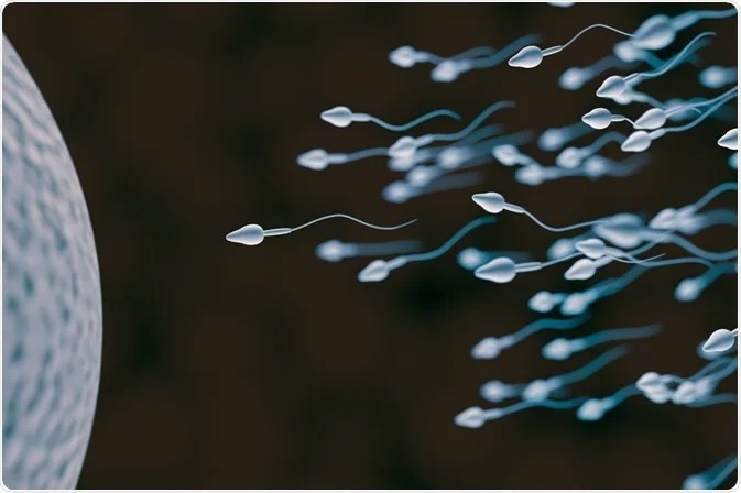
Our research focuses on the characterization of the molecular mechanisms which govern sperm flagellum assembly as well as sperm motility and fertilization potential. In parallel, we are interested in defining the genetic causes and patho-physiological mechanisms associated with human male infertility mainly due to asthenozoospermia, with the aim of improving current procedures of assisted reproduction technologies and developing novel therapeutics.

Research Topics
SPERM SIGNALING & ENERGY METABOLIC PATHWAYS
Deciphering the signalling and metabolic pathways required for sperm motility and fertilization potential.
SPERM PATHOPHYSIOLOGY
Defining the genetic causes and molecular mechanisms associated with male infertility due to asthenozoospermia.
The TEAM

Aminata TOURE, PI
Aminata Touré was born in Paris and obtained her PhD in 2000 under the supervision of Dr Gérard Gacon (University Paris Descartes, France), identifying and characterizing novel proteins involved in Rho GTPases signaling pathways and expressed in human and mouse spermatogenic cells.
Following her PhD, she joined the laboratory of Dr Paul Burgoyne & Dr Robin Lovell-Badge at the National Institute for Medical Research in London (MRC, London, UK) to work on the characterization of multi-copy gene families located on the mouse Y chromosome. Her work was pioneer in demonstrating that the Y long arm chromosome encode for proteins that are essential for mouse spermiogenesis and fertility.
In 2004, she was recruited at the Institut Cochin (Paris, France) as a CNRS research fellow (CR2) to develop her program in the field of ion channels and sperm physiology. From then, she has been using cell biology, molecular biology, biochemistry and mouse genetics to define the molecular mechanisms associated with sperm motility and fertilization potential. In particular, her work highlighted the role of SLC26 and CFTR anion channels in sperm motility and capacitation. In 2013, she was promoted CNRS Research Director (DR2) and extended her research program to the genetics and pathophysiology of male infertility mainly due to asthenozoospermia.
In 2020, she joined the Institute for Advanced Biosciences (Grenoble, France) where she is now leading the team “Physiology and Physiopathology of Sperm Cells” and piloting novel research programs in the field of sperm signaling and metabolic pathways.
Joao RIBEIRO

Joao RIBEIRO, Post-doctoral Fellow
Lauryne BRY

Lauryne BRY, Engineer Assistant (CDD)
Ismail MOUSSAOUI
Ismail MOUSSAOUI, Engineer Assistant (CDD)
Sarah LIBERT-BAREC

Sarah LIBERT-BAREC, Research Engineer (CDD)
Pablo RAMIREZ

Pablo RAMIREZ, Erasmus student
Marjorie WHITFIELD
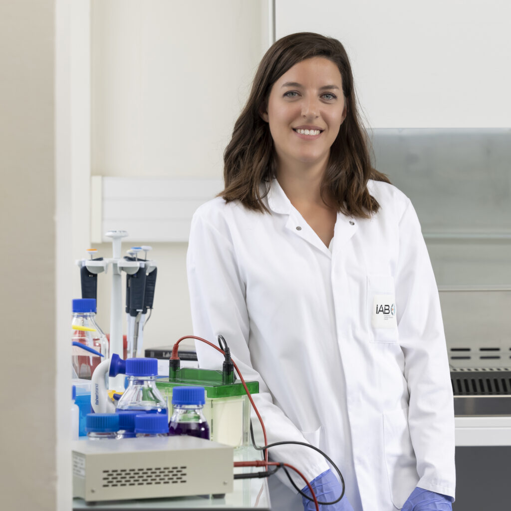
Marjorie WHITFIELD, Researcher (Inserm CRCN)
During my Ph.D training (completed in 2017) at the GReD laboratory (University Clermont-Auvergne, Clermont Ferrand) under the supervision of Dr Fabrice Saez and Pr Joël Drevet, I have studied the physiology of spermatozoa with a particular interest in understanding their functional maturation during the transit in the epididymis and the female genital tract. Following this work, I joined the research group of Dr. Aminata Touré at the Cochin Institute (University of Paris), where I focused on the genetic causes and pathophysiological mechanisms that are associated with human asthenozoospermia. Within different consortia including geneticists and physicians in reproductive biology, my work contributed to the characterization of several gene mutations affecting the composition and assembly of axonemal dynein arm complexes, which are critical for cilia and flagella beating.
Since 2021, I have joined the Institute for Advanced Biosciences (Grenoble) to develop my own research program on the biogenesis of sperm flagellum. My long-term goal is to decipher the molecular and cellular mechanisms sustaining sperm flagellum assembly and maintenance, and to assess the contribution of the annulus in these processes. To conduct my research program, I have established over the past years several strong collaborations with national and international leaders in the field of genetics, ciliopathies and reproductive biology.
Along my research activities, I am actively participating in science communication through different social media. In 2021, I also received the Young Talent L’Oréal-Unesco Foundation Prize “For Women In Science” for my work on male reproductive biology and infertility, an award which aims at promoting women’s careers in science. This prize constitutes an important foundation to initiate my research program regarding the biogenesis of sperm flagellum.
Violaine SIMON

Violaine SIMON, Lecturer
Maëva DROUAULT

Maëva DROUAULT, Post-doctoral Fellow
After a Master degree in Molecular Genetics completed at the University of Bordeaux, I obtained my PhD in Molecular and Cellular Biology at the University of Caen Normandy. My thesis conducted under the supervision of Dr Christelle Delalande and Dr Vincent Hanoux, was focused on Male Reproductive Biology and Toxicology. In particular I have investigated the role of estrogen signaling in rat testis and its alteration by endocrine disruptors.
In March 2023, I have joined the research group of Dr Aminata Touré at the Institute for Advanced Biosciences (Grenoble) to work on sperm motility and metabolism as a postdoctoral researcher. I am particularly involved in the SPERMetabo project funded by the ANR, which aims at deciphering the metabolic pathways supporting energy production in human and mouse sperm cells, and at characterizing the consequences of their deficiencies in the context of human male infertility.
Alumni
Elma El Khouri (2013-2016)
Charles Coutton (2017)
Marjorie Whitfield (2018-2022)
Pierre Lhuillier (2004-2008)
Baptiste Rode (2007-2011)
Thassadite Dirami (2009-2012)
Emma Cavarocchi (2019-2023)
Emma Cavarocchi Italian born, I obtained a Bachelor’s Degree in Biotechnologies at the University of Bologna (Italy). During my first internship, I undertook the caracterization of the Caretta caretta turtle microbiota in the Laboratory of Microbiology of the Unibo Pharmacy and Biotechnologies Department.
I then attended the Master 1 in Molecular Biology at the University of Padova (Italy) and had the opportunity to join a Double Degree program moving to the Paris Diderot University in France (Magistère Européen de Génétique – Master 2). During this cursus, I spent 6 months in the laboratory of Genomics, Epigenetics and Physiopathology of Reproduction at the Institut Cochin, working on human asthenozoospermia under the supervision of Dr Aminata Touré.
France definitely became my second home, as in October 2019, on the trail of my Master 2 internship, I started a PhD to better explore the genetics and pathophysiology of asthenozoospermia. In 2020 I followed my supervisor Aminata Touré moving to the Institute for Advanced Biosciences in Grenoble. I obtained the PhD degree at the University Paris Cité on January 23rd 2023.
Apart from being fascinated by the biological mechanisms underlying reproduction, I like spending my time with a book, a good movie and an apèro with my friends. I also love exploring nature, cultures and food!
Baptiste Rode (2006)
Thassadite Dirami (2008)
Nathalie Da Silva (2011)
Maelle Givelet (2013)
Zeinab Sakheli (2014)
Marhaba Chaudhry (2015)
Precilia Homand (2015)
Jean-Fabrice Nsota Mbango (2016)
Julie Tek (2016)
Emma Cavarocchi (2019)
Coraline Joly (2022)
Fadwa Jreijiri (2022)
Abderazak Sidi Salah (2023)
Oriane Huet (2024)
Sébastien Pichon (2006)
Thassadite Dirami (2007)
Audrey Chansard (2010)
Lucie Puron (2011)
Rabah Tamoumi (2012)
Justine LOUDAN (2023)
Elsie Billon (2024)
Charlotte Dussaix (2024)
Publications
All / 2020-2025 / 2020-2024 / 2020-2023 / 2020-2022 / 2020-2021 / 2015-2019 / 2010-2014 / 2005-2009 / 1995-2004
Human asthenozoospermia: Update on genetic causes, patient management, and clinical strategies.
Emma Cavarocchi, Drouault M, Ribeiro JC; Simon V Marjorie Whitfield, and Aminata TOURE
Andrology , (2025)
CCDC65, encoding a component of the axonemal Nexin-Dynein regulatory complex, is required for sperm flagellum structure in humans.
Jreijiri F, Cavarocchi E, Amiri-Yekta A, Cazin C, Hosseini SH, El Khouri E, Patrat C, Thierry-Mieg N, Ray PF, Dulioust E, Whitfield M, Touré A
Clinical Genetics , (2024)
The annulus: composition, role and importance in sperm flagellum biogenesis and male fertility
Marjorie Whitfield
Basic and clinical andrology , (2024)
DNALI1 interacts with the MEIG1/PACRG complex within the manchette and is required for proper sperm flagellum assembly in mice
Yap, Yi Tian and Li, Wei and Huang, Qian and Zhou, Qi and Zhang, David and Sheng, Yi and Mladenovic-Lucas, Ljljiana and Yee, Siu-Pok and Orwig, Kyle E and Granneman, James G and Williams, David C and Hess, Rex A and Touré, Aminata and Zhang, Zhibing
eLife 12, e79620 (2023)
DNALI1 interacts with the MEIG1/PACRG complex within the manchette and is required for proper sperm flagellum assembly in mice
The manchette is a transient and unique structure present in elongating spermatids and required for proper differentiation of the germ cells during spermatogenesis. Previous work indicated that the MEIG1/PACRG complex locates in the manchette and is involved in the transport of cargos, such as SPAG16L, to build the sperm flagellum. Here, using co-immunoprecipitation and pull-down approaches in various cell systems, we established that DNALI1, an axonemal component originally cloned from <i>Chlamydomonas reinhardtii</i>, recruits and stabilizes PACRG and we confirm in vivo, the co-localization of DNALI1 and PACRG in the manchette by immunofluorescence of elongating murine spermatids. We next generated mice with a specific deficiency of DNALI1 in male germ cells, and observed a dramatic reduction of the sperm cells, which results in male infertility. In addition, we observed that the majority of the sperm cells exhibited abnormal morphology including misshapen heads, bent tails, enlarged midpiece, discontinuous accessory structure, emphasizing the importance of DNALI1 in sperm differentiation. Examination of testis histology confirmed impaired spermiogenesis in the mutant mice. Importantly, while testicular levels of MEIG1, PACRG, and SPAG16L proteins were unchanged in the <i>Dnali1</i> mutant mice, their localization within the manchette was greatly affected, indicating that DNALI1 is required for the formation of the MEIG1/PACRG complex within the manchette. Interestingly, in contrast to MEIG1 and PACRG-deficient mice, the DNALI1-deficient mice also showed impaired sperm spermiation/individualization, suggesting additional functions beyond its involvement in the manchette structure. Overall, our work identifies DNALI1 as a protein required for sperm development.
Identification of IQCH as a calmodulin-associated protein required for sperm motility in humans
Emma Cavarocchi and Camille Sayou and Patrick Lorès and Caroline Cazin and Laurence Stouvenel and Elma El Khouri and Charles Coutton and Zine-Eddine Kherraf and Catherine Patrat and Jérôme Govin and Nicolas Thierry-Mieg and Marjorie Whitfield and Pierre F. Ray and Emmanuel Dulioust and Aminata Touré
iScience 26, 107354 (2023)
Identification of IQCH as a calmodulin-associated protein required for sperm motility in humans
SummarySperm fertilization ability mainly relies on proper sperm progression through the female genital tract and capacitation, which involves phosphorylation signaling pathways triggered by calcium and bicarbonate. We performed exome sequencing of an infertile asthenozoospermic patient and identified truncating variants in MAP7D3, encoding a microtubule-associated protein, and IQCH, encoding a protein of unknown function with enzymatic and signaling features. We demonstrate the deleterious impact of both variants on sperm transcripts and proteins from the patient. We show that, in vitro, patient spermatozoa could not induce the phosphorylation cascades associated with capacitation. We also provide evidence for IQCH association with calmodulin, a well-established calcium-binding protein that regulates the calmodulin kinase. Notably, we describe IQCH spatial distribution around the sperm axoneme, supporting its function within flagella. Overall, our work highlights the cumulative pathological impact of gene mutations and identifies IQCH as a key protein required for sperm motility and capacitation.
CCDC65, encoding a component of the axonemal Nexin-Dynein regulatory complex, is required for sperm flagellum structure in humans
Fadwa Jreijiri, Emma Cavarocchi, Amir Amiri-Yekta, Caroline Cazin, Seyedeh-Hanieh Hosseini, Elma El Khouri, Catherine Patrat, Nicolas Thierry-Mieg, Pierre F Ray, Emmanuel Dulioust, Marjorie Whitfield, Aminata Touré
Clinical Genetics , (2023)
CCDC65, encoding a component of the axonemal Nexin-Dynein regulatory complex, is required for sperm flagellum structure in humans
Sperm flagella share an evolutionary conserved microtubule-based structure with motile cilia expressed at the surface of several cell types, such as the airways epithelial cells. As a result, male infertility can be observed as an isolated condition or a syndromic trait, illustrated by Primary Cilia Dyskinesia (PCD). We report two unrelated patients showing multiple morphological abnormalities of the sperm flagella (MMAF) and carrying distinct homozygous truncating variants in the PCD-associated gene CCDC65. We characterized one of the identified variants (c.1208del; p.Asn403Ilefs*9), which induces the near absence of CCDC65 protein in patient sperm. In Chlamydomonas, CCDC65 ortholog (DRC2, FAP250) is a component of the Nexin-Dynein Regulatory complex (N-DRC), which interconnects microtubule doublets and coordinates dynein arms activity. In sperm cells from the patient, we also show the loss of GAS8, another component of the N-DRC, supporting a structural/functional link between the two proteins. Our work indicates that, similarly to ciliary axoneme, CCDC65 is required for sperm flagellum structure. Importantly, our work provides first evidence that mutations in the PCD-associated gene CCDC65 also cause asthenozoospermia.
Novel axonemal protein ZMYND12 interacts with TTC29 and DNAH1, and is required for male fertility and flagellum function.
Dacheux D, Martinez G, Broster Reix CE, Beurois J, Lores P, Tounkara M, Dupuy JW, Robinson DR, Loeuillet C, Lambert E, Wehbe Z, Escoffier J, Coutton C
ELife , (2023)
Results and perinatal outcomes from 189 ICSI cycles of couples with asthenozoospermic men and flagellar defects assessed by transmission electron microscopy.
Boursier A, Boudry A, Mitchell V, Loyens A, Rives N, Moerman A, Thomas L, Escudier E, Toure A, Whitfield M, Coutton C, Martinez G, Ray PF, Barbotin AL
Reproductive Biomedicine Online , (2023)
Identification of IQCH as a calmodulin-associated protein required for sperm motility in humans.
Cavarocchi E, Sayou C, Lorès P, Cazin C, Stouvenel L, El Khouri E, Coutton C, Kherraf ZE, Patrat C, Govin J, Thierry-Mieg N, Whitfield M, Ray PF, Touré A
IScience , (2023)
Sperm Ion Transporters and Channels in Human Asthenozoospermia: Genetic Etiology, Lessons from Animal Models, and Clinical Perspectives
Cavarocchi, Emma and Whitfield, Marjorie and Saez, Fabrice and Touré, Aminata
International Journal of Molecular Sciences 23, (2022)
Sperm Ion Transporters and Channels in Human Asthenozoospermia: Genetic Etiology, Lessons from Animal Models, and Clinical Perspectives
In mammals, sperm fertilization potential relies on efficient progression within the female genital tract to reach and fertilize the oocyte. This fundamental property is supported by the flagellum, an evolutionarily conserved organelle that provides the mechanical force for sperm propulsion and motility. Importantly several functional maturation events that occur during the journey of the sperm cells through the genital tracts are necessary for the activation of flagellar beating and the acquisition of fertilization potential. Ion transporters and channels located at the surface of the sperm cells have been demonstrated to be involved in these processes, in particular, through the activation of downstream signaling pathways and the promotion of novel biochemical and electrophysiological properties in the sperm cells. We performed a systematic literature review to describe the currently known genetic alterations in humans that affect sperm ion transporters and channels and result in asthenozoospermia, a pathophysiological condition defined by reduced or absent sperm motility and observed in nearly 80% of infertile men. We also present the physiological relevance and functional mechanisms of additional ion channels identified in the mouse. Finally, considering the state-of-the art, we discuss future perspectives in terms of therapeutics of asthenozoospermia and male contraception.
Genetic diagnosis, sperm phenotype and ICSI outcome in case of severe asthenozoospermia with multiple morphological abnormalities of the flagellum
Ferreux, Lucile and Bourdon, Mathilde and Chargui, Ahmed and Schmitt, Alain and Stouvenel, Laurence and Lorès, Patrick and Ray, Pierre and Lousqui, Johanna and Pocate-Cheriet, Khaled and Santulli, Pietro and Dulioust, Emmanuel and Touré, Aminata and Patrat, Catherine
Human reproduction (Oxford, England) , (2021)
Genetic diagnosis, sperm phenotype and ICSI outcome in case of severe asthenozoospermia with multiple morphological abnormalities of the flagellum
Study question
Are ICSI outcomes impaired in cases of severe asthenozoospermia with multiple morphological abnormalities of the flagellum (MMAF phenotype)?
Summary answer
Despite occasional technical difficulties, ICSI outcomes for couples with MMAF do not differ from those of other couples requiring ICSI, irrespective of the genetic defect.
What is known already
Severe asthenozoospermia, especially when associated with the MMAF phenotype, results in male infertility. Recent findings have confirmed that a genetic aetiology is frequently responsible for this phenotype. In such situations, pregnancies can be achieved using ICSI. However, few studies to date have provided detailed analyses regarding the flagellar ultrastructural defects underlying this phenotype, its genetic aetiologies, and the results of ICSI in such cases of male infertility.
Study design, size, duration
We performed a retrospective study of 25 infertile men exhibiting severe asthenozoospermia associated with the MMAF phenotype identified through standard semen analysis. They were recruited at an academic centre for assisted reproduction in Paris (France) between 2009 and 2017. Transmission electron microscopy (TEM) and whole exome sequencing (WES) were performed in order to determine the sperm ultrastructural phenotype and the causal mutations, respectively. Finally 20 couples with MMAF were treated by assisted reproductive technologies based on ICSI.
Participants/materials, setting, methods
Patients with MMAF were recruited based on reduced sperm progressive motility and increased frequencies of absent, short, coiled or irregular flagella compared with those in sperm from fertile control men. A quantitative analysis of the several ultrastructural defects was performed for the MMAF patients and for fertile men. The ICSI results obtained for 20 couples with MMAF were compared to those of 378 men with oligoasthenoteratozoospermia but no MMAF as an ICSI control group.
Main results and the role of chance
TEM analysis and categorisation of the flagellar anomalies found in these patients provided important information regarding the structural defects underlying asthenozoospermia and sperm tail abnormalities. In particular, the absence of the central pair of axonemal microtubules was the predominant anomaly observed more frequently than in control sperm (P < 0.01). Exome sequencing, performed for 24 of the 25 patients, identified homozygous or compound heterozygous pathogenic mutations in CFAP43, CFAP44, CFAP69, DNAH1, DNAH8, AK7, TTC29 and MAATS1 in 13 patients (54.2%) (11 affecting MMAF genes and 2 affecting primary ciliary dyskinesia (PCD)-associated genes). A total of 40 ICSI cycles were undertaken for 20 MMAF couples, including 13 cycles (for 5 couples) where a hypo-osmotic swelling (HOS) test was required due to absolute asthenozoospermia. The fertilisation rate was not statistically different between the MMAF (65.7%) and the non-MMAF (66.0%) couples and it did not differ according to the genotype or the flagellar phenotype of the subjects or use of the HOS test. The clinical pregnancy rate per embryo transfer did not differ significantly between the MMAF (23.3%) and the non-MMAF (37.1%) groups. To date, 7 of the 20 MMAF couples have achieved a live birth from the ICSI attempts, with 11 babies born without any birth defects.
Limitations, reasons for caution
The ICSI procedure outcomes were assessed retrospectively on a small number of affected subjects and should be confirmed on a larger cohort. Moreover, TEM analysis could not be performed for all patients due to low sperm concentrations, and WES results are not yet available for all of the included men.
Wider implications of the findings
An early and extensive phenotypic and genetic investigation should be considered for all men requiring ICSI for severe asthenozoospermia. Although our study did not reveal any adverse ICSI outcomes associated with MMAF, we cannot rule out that some rare genetic causes could result in low fertilisation or pregnancy rates.
Study funding/competing interest(s)
No external funding was used for this study and there are no competing interests.
Trial registration number
N/A.
Bi-allelic truncating variants in CFAP206 cause male infertility in human and mouse
Shen, Qunshan and Martinez, Guillaume and Liu, Hongbin and Beurois, Julie and Wu, Huan and Amiri-Yekta, Amir and Liang, Dan and Kherraf, Zine-Eddine and Bidart, Marie and Cazin, Caroline and Celse, Tristan and Satre, Véronique and Thierry-Mieg, Nicolas and Whitfield, Marjorie and Touré, Aminata and Song, Bing and Lv, Mingrong and Li, Kuokuo and Liu, Chunyu and Tao, Fangbiao and He, Xiaojin and Zhang, Feng and Arnoult, Christophe and Ray, Pierre F and Cao, Yunxia and Coutton, Charles
Human genetics 140, 1367—1377 (2021)
Bi-allelic truncating variants in CFAP206 cause male infertility in human and mouse
Spermatozoa are polarized cells with a head and a flagellum joined together by the connecting piece. Flagellum integrity is critical for normal sperm function, and flagellum defects consistently lead to male infertility. Multiple morphological abnormalities of the flagella (MMAF) is a distinct sperm phenotype consistently leading to male infertility due to a reduced or absent sperm motility associated with severe morphological and ultrastructural flagellum defects. Despite numerous genes recently described to be recurrently associated with MMAF, more than half of the cases analyzed remain unresolved, suggesting that many yet uncharacterized gene defects account for this phenotype. By performing a retrospective exome analysis of the unsolved cases from our initial cohort of 167 infertile men with a MMAF phenotype, we identified one individual carrying a homozygous frameshift variant in CFAP206, a gene encoding a microtubule-docking adapter for radial spoke and inner dynein arm. Immunostaining experiments in the patient’s sperm cells demonstrated the absence of WDR66 and RSPH1 proteins suggesting severe radial spokes and calmodulin and spoke-associated complex defects. Using the CRISPR-Cas9 technique, we generated homozygous Cfap206 knockout (KO) mice which presented with male infertility due to functional, structural and ultrastructural sperm flagellum defects associated with a very low rate of embryo development using ICSI. Overall, we showed that CFAP206 is essential for normal sperm flagellum structure and function in human and mouse and that bi-allelic mutations in CFAP206 cause male infertility in man and mouse by inducing morphological and functional defects of the sperm flagellum that may also cause ICSI failures.
Correction to: A missense mutation in IFT74, encoding for an essential component for intraflagellar transport of Tubulin, causes asthenozoospermia and male infertility without clinical signs of Bardet-Biedl syndrome
Lorès, Patrick and Kherraf, Zine-Eddine and Amiri-Yekta, Amir and Whitfield, Marjorie and Daneshipour, Abbas and Stouvenel, Laurence and Cazin, Caroline and Cavarocchi, Emma and Coutton, Charles and Llabador, Marie-Astrid and Arnoult, Christophe and Thierry-Mieg, Nicolas and Ferreux, Lucile and Patrat, Catherine and Hosseini, Seyedeh-Hanieh and Mustapha, Selima Fourati Ben and Zouari, Raoudha and Dulioust, Emmanuel and Ray, Pierre F and Touré, Aminata
Human genetics 140, 1045 (2021)
A missense mutation in IFT74, encoding for an essential component for intraflagellar transport of Tubulin, causes asthenozoospermia and male infertility without clinical signs of Bardet-Biedl syndrome
Lorès, Patrick and Kherraf, Zine-Eddine and Amiri-Yekta, Amir and Whitfield, Marjorie and Daneshipour, Abbas and Stouvenel, Laurence and Cazin, Caroline and Cavarocchi, Emma and Coutton, Charles and Llabador, Marie-Astrid and Arnoult, Christophe and Thierry-Mieg, Nicolas and Ferreux, Lucile and Patrat, Catherine and Hosseini, Seyedeh-Hanieh and Mustapha, Selima Fourati Ben and Zouari, Raoudha and Dulioust, Emmanuel and Ray, Pierre F and Touré, Aminata
Human genetics 140, 1031—1043 (2021)
A missense mutation in IFT74, encoding for an essential component for intraflagellar transport of Tubulin, causes asthenozoospermia and male infertility without clinical signs of Bardet-Biedl syndrome
Cilia and flagella are formed around an evolutionary conserved microtubule-based axoneme and are required for fluid and mucus clearance, tissue homeostasis, cell differentiation and movement. The formation and maintenance of cilia and flagella require bidirectional transit of proteins along the axonemal microtubules, a process called intraflagellar transport (IFT). In humans, IFT defects contribute to a large group of systemic diseases, called ciliopathies, which often display overlapping phenotypes. By performing exome sequencing of a cohort of 167 non-syndromic infertile men displaying multiple morphological abnormalities of the sperm flagellum (MMAF) we identified two unrelated patients carrying a homozygous missense variant adjacent to a splice donor consensus site of IFT74 (c.256G > A;p.Gly86Ser). IFT74 encodes for a core component of the IFT machinery that is essential for the anterograde transport of tubulin. We demonstrate that this missense variant affects IFT74 mRNA splicing and induces the production of at least two distinct mutant proteins with abnormal subcellular localization along the sperm flagellum. Importantly, while IFT74 deficiency was previously implicated in two cases of Bardet-Biedl syndrome, a pleiotropic ciliopathy with variable expressivity, our data indicate that this missense mutation only results in primary male infertility due to MMAF, with no other clinical features. Taken together, our data indicate that the nature of the mutation adds a level of complexity to the clinical manifestations of ciliary dysfunction, thus contributing to the expanding phenotypical spectrum of ciliopathies.
Identification and Characterization of the Most Common Genetic Variant Responsible for Acephalic Spermatozoa Syndrome in Men Originating from North Africa
Cazin, Caroline and Boumerdassi, Yasmine and Martinez, Guillaume and Fourati Ben Mustapha, Selima and Whitfield, Marjorie and Coutton, Charles and Thierry-Mieg, Nicolas and Di Pizio, Pierre and Rives, Nathalie and Arnoult, Christophe and Touré, Aminata and Ray, Pierre F and Zouari, Raoudha and Sifer, Christophe and Kherraf, Zine-Eddine
International journal of molecular sciences 22, (2021)
Identification and Characterization of the Most Common Genetic Variant Responsible for Acephalic Spermatozoa Syndrome in Men Originating from North Africa
Acephalic spermatozoa syndrome (ASS) is a rare but extremely severe type of teratozoospermia, defined by the presence of a majority of headless flagella and a minority of tail-less sperm heads in the ejaculate. Like the other severe monomorphic teratozoospermias, ASS has a strong genetic basis and is most often caused by bi-allelic variants in SUN5 (Sad1 and UNC84 domain-containing 5). Using whole exome sequencing (WES), we investigated a cohort of nine infertile subjects displaying ASS. These subjects were recruited in three centers located in France and Tunisia, but all originated from North Africa. Sperm from subjects carrying candidate genetic variants were subjected to immunofluorescence analysis and transmission electron microscopy. Moreover, fluorescent in situ hybridization (FISH) was performed on sperm nuclei to assess their chromosomal content. Variant filtering permitted us to identify the same SUN5 homozygous frameshift variant (c.211+1_211+2dup) in 7/9 individuals (78%). SUN5 encodes a protein localized on the posterior part of the nuclear envelope that is necessary for the attachment of the tail to the sperm head. Immunofluorescence assays performed on sperm cells from three mutated subjects revealed a total absence of SUN5, thus demonstrating the deleterious impact of the identified variant on protein expression. Transmission electron microscopy showed a conserved flagellar structure and a slightly decondensed chromatin. FISH did not highlight a higher rate of chromosome aneuploidy in spermatozoa from SUN5 patients compared to controls, indicating that intra-cytoplasmic sperm injection (ICSI) can be proposed for patients carrying the c.211+1_211+2dup variant. These results suggest that the identified SUN5 variant is the main cause of ASS in the North African population. Consequently, a simple and inexpensive genotyping of the 211+1_211+2dup variant could be beneficial for affected men of North African origin before resorting to more exhaustive genetic analyses.
Deleterious variants in X-linked CFAP47 induce asthenoteratozoospermia and primary male infertility
Liu, Chunyu and Tu, Chaofeng and Wang, Lingbo and Wu, Huan and Houston, Brendan J and Mastrorosa, Francesco K and Zhang, Wen and Shen, Ying and Wang, Jiaxiong and Tian, Shixiong and Meng, Lanlan and Cong, Jiangshan and Yang, Shenmin and Jiang, Yiwen and Tang, Shuyan and Zeng, Yuyan and Lv, Mingrong and Lin, Ge and Li, Jinsong and Saiyin, Hexige and He, Xiaojin and Jin, Li and Touré, Aminata and Ray, Pierre F and Veltman, Joris A and Shi, Qinghua and O’Bryan, Moira K and Cao, Yunxia and Tan, Yue-Qiu and Zhang, Feng
American journal of human genetics 108, 309—323 (2021)
Deleterious variants in X-linked CFAP47 induce asthenoteratozoospermia and primary male infertility
Asthenoteratozoospermia characterized by multiple morphological abnormalities of the flagella (MMAF) has been identified as a sub-type of male infertility. Recent progress has identified several MMAF-associated genes with an autosomal recessive inheritance in human affected individuals, but the etiology in approximately 40% of affected individuals remains unknown. Here, we conducted whole-exome sequencing (WES) and identified hemizygous missense variants in the X-linked CFAP47 in three unrelated Chinese individuals with MMAF. These three CFAP47 variants were absent in human control population genome databases and were predicted to be deleterious by multiple bioinformatic tools. CFAP47 encodes a cilia- and flagella-associated protein that is highly expressed in testis. Immunoblotting and immunofluorescence assays revealed obviously reduced levels of CFAP47 in spermatozoa from all three men harboring deleterious missense variants of CFAP47. Furthermore, WES data from an additional cohort of severe asthenoteratozoospermic men originating from Australia permitted the identification of a hemizygous Xp21.1 deletion removing the entire CFAP47 gene. All men harboring hemizygous CFAP47 variants displayed typical MMAF phenotypes. We also generated a Cfap47-mutated mouse model, the adult males of which were sterile and presented with reduced sperm motility and abnormal flagellar morphology and movement. However, fertility could be rescued by the use of intra-cytoplasmic sperm injections (ICSIs). Altogether, our experimental observations in humans and mice demonstrate that hemizygous mutations in CFAP47 can induce X-linked MMAF and asthenoteratozoospermia, for which good ICSI prognosis is suggested. These findings will provide important guidance for genetic counseling and assisted reproduction treatments.
The sodium/proton exchanger SLC9C1 (sNHE) is essential for human sperm motility and fertility
Cavarocchi, Emma and Whitfield, Marjorie and Chargui, Ahmed and Stouvenel, Laurence and Lorès, Patrick and Coutton, Charles and Arnoult, Christophe and Santulli, Pietro and Patrat, Catherine and Thierry-Mieg, Nicolas and Ray, Pierre F and Dulioust, Emmanuel and Touré, Aminata
Clinical genetics 99, 684—693 (2021)
The sodium/proton exchanger SLC9C1 (sNHE) is essential for human sperm motility and fertility
Asthenozoospermia, defined by the absence or reduction of sperm motility, constitutes the most frequent cause of human male infertility. This pathological condition is caused by morphological and/or functional defects of the sperm flagellum, which preclude proper sperm progression. While in the last decade many causal genes were identified for asthenozoospermia associated with severe sperm flagellar defects, the causes of purely functional asthenozoospermia are still poorly defined. We describe here the case of an infertile man, displaying asthenozoospermia without major morphological flagellar anomalies and carrying a homozygous splicing mutation in SLC9C1 (sNHE), which we identified by whole-exome sequencing. SLC9C1 encodes a sperm-specific sodium/proton exchanger, which in mouse regulates pH homeostasis and interacts with the soluble adenylyl cyclase (sAC), a key regulator of the signalling pathways involved in sperm motility and capacitation. We demonstrate by means of RT-PCR, immunodetection and immunofluorescence assays on patient’s semen samples that the homozygous splicing mutation (c.2748 + 2 T > C) leads to in-frame exon skipping resulting in a deletion in the cyclic nucleotide-binding domain of the protein. Our work shows that in human, similar to mouse, SLC9C1 is required for sperm motility. Overall, we establish a homozygous truncating mutation in SLC9C1 as a novel cause of human asthenozoospermia and infertility.
Tubulin glycylation controls axonemal dynein activity, flagellar beat, and male fertility
Gadadhar, Sudarshan and Alvarez Viar, Gonzalo and Hansen, Jan Niklas and Gong, An and Kostarev, Aleksandr and Ialy-Radio, Côme and Leboucher, Sophie and Whitfield, Marjorie and Ziyyat, Ahmed and Touré, Aminata and Alvarez, Luis and Pigino, Gaia and Janke, Carsten
Science (New York, N.Y.) 371, (2021)
Tubulin glycylation controls axonemal dynein activity, flagellar beat, and male fertility
Posttranslational modifications of the microtubule cytoskeleton have emerged as key regulators of cellular functions, and their perturbations have been linked to a growing number of human pathologies. Tubulin glycylation modifies microtubules specifically in cilia and flagella, but its functional and mechanistic roles remain unclear. In this study, we generated a mouse model entirely lacking tubulin glycylation. Male mice were subfertile owing to aberrant beat patterns of their sperm flagella, which impeded the straight swimming of sperm cells. Using cryo-electron tomography, we showed that lack of glycylation caused abnormal conformations of the dynein arms within sperm axonemes, providing the structural basis for the observed dysfunction. Our findings reveal the importance of microtubule glycylation for controlled flagellar beating, directional sperm swimming, and male fertility.
The genetic architecture of morphological abnormalities of the sperm tail
Touré, Aminata and Martinez, Guillaume and Kherraf, Zine-Eddine and Cazin, Caroline and Beurois, Julie and Arnoult, Christophe and Ray, Pierre F and Coutton, Charles
Human genetics 140, 21—42 (2021)
The genetic architecture of morphological abnormalities of the sperm tail
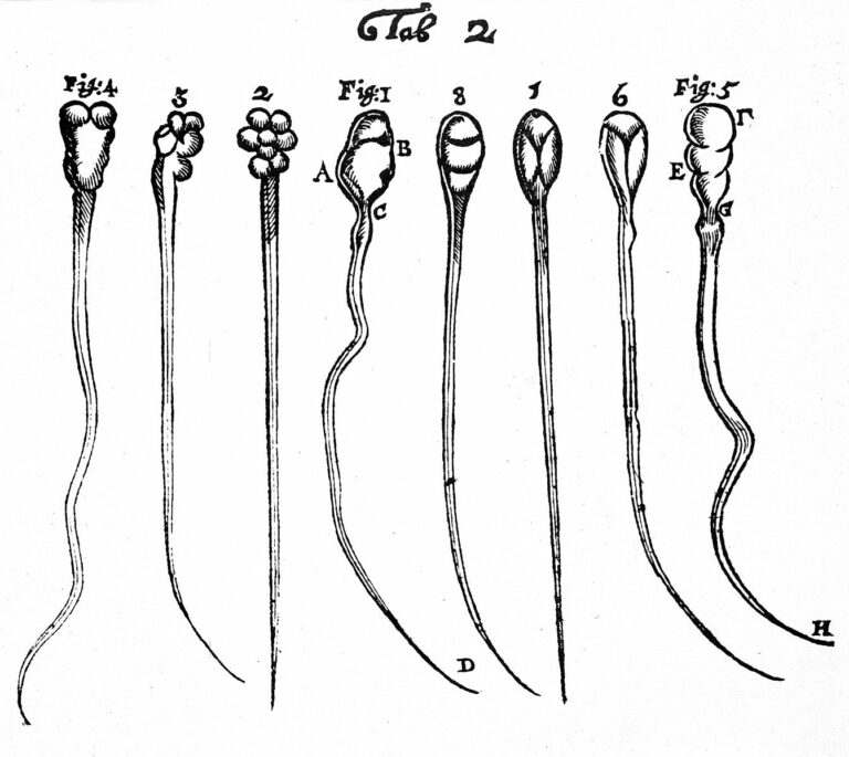
Spermatozoa contain highly specialized structural features reflecting unique functions required for fertilization. Among them, the flagellum is a sperm-specific organelle required to generate the motility, which is essential to reach the egg. The flagellum integrity is, therefore, critical for normal sperm function and flagellum defects consistently lead to male infertility due to reduced or absent sperm motility defined as asthenozoospermia. Multiple morphological abnormalities of the flagella (MMAF), also called short tails, is among the most severe forms of sperm flagellum defects responsible for male infertility and is characterized by the presence in the ejaculate of spermatozoa being short, coiled, absent and of irregular caliber. Recent studies have demonstrated that MMAF is genetically heterogeneous which is consistent with the large number of proteins (over one thousand) localized in the human sperm flagella. In the past 5 years, genomic investigation of the MMAF phenotype allowed the identification of 18 genes whose mutations induce MMAF and infertility. Here we will review information about those genes including their expression pattern, the features of the encoded proteins together with their localization within the different flagellar protein complexes (axonemal or peri-axonemal) and their potential functions. We will categorize the identified MMAF genes following the protein complexes, functions or biological processes they may be associated with, based on the current knowledge in the field.
{Genetic diagnosis, sperm phenotype and ICSI outcome in case of severe asthenozoospermia with multiple morphological abnormalities of the flagellum}
Ferreux, Lucile and Bourdon, Mathilde and Chargui, Ahmed and Schmitt, Alain and Stouvenel, Laurence and Lorès, Patrick and Ray, Pierre and Lousqui, Johanna and Pocate-Cheriet, Khaled and Santulli, Pietro and Dulioust, Emmanuel and Toure, Aminata and Patrat, Catherine
Human Reproduction 36, 2848-2860 (2021)
{Genetic diagnosis, sperm phenotype and ICSI outcome in case of severe asthenozoospermia with multiple morphological abnormalities of the flagellum}
{Are ICSI outcomes impaired in cases of severe asthenozoospermia with multiple morphological abnormalities of the flagellum (MMAF phenotype)?Despite occasional technical difficulties, ICSI outcomes for couples with MMAF do not differ from those of other couples requiring ICSI, irrespective of the genetic defect.Severe asthenozoospermia, especially when associated with the MMAF phenotype, results in male infertility. Recent findings have confirmed that a genetic aetiology is frequently responsible for this phenotype. In such situations, pregnancies can be achieved using ICSI. However, few studies to date have provided detailed analyses regarding the flagellar ultrastructural defects underlying this phenotype, its genetic aetiologies, and the results of ICSI in such cases of male infertility.We performed a retrospective study of 25 infertile men exhibiting severe asthenozoospermia associated with the MMAF phenotype identified through standard semen analysis. They were recruited at an academic centre for assisted reproduction in Paris (France) between 2009 and 2017. Transmission electron microscopy (TEM) and whole exome sequencing (WES) were performed in order to determine the sperm ultrastructural phenotype and the causal mutations, respectively. Finally 20 couples with MMAF were treated by assisted reproductive technologies based on ICSI.Patients with MMAF were recruited based on reduced sperm progressive motility and increased frequencies of absent, short, coiled or irregular flagella compared with those in sperm from fertile control men. A quantitative analysis of the several ultrastructural defects was performed for the MMAF patients and for fertile men. The ICSI results obtained for 20 couples with MMAF were compared to those of 378 men with oligoasthenoteratozoospermia but no MMAF as an ICSI control group.TEM analysis and categorisation of the flagellar anomalies found in these patients provided important information regarding the structural defects underlying asthenozoospermia and sperm tail abnormalities. In particular, the absence of the central pair of axonemal microtubules was the predominant anomaly observed more frequently than in control sperm (P \\< 0.01). Exome sequencing, performed for 24 of the 25 patients, identified homozygous or compound heterozygous pathogenic mutations in CFAP43, CFAP44, CFAP69, DNAH1, DNAH8, AK7, TTC29 and MAATS1 in 13 patients (54.2\\%) (11 affecting MMAF genes and 2 affecting primary ciliary dyskinesia (PCD)-associated genes). A total of 40 ICSI cycles were undertaken for 20 MMAF couples, including 13 cycles (for 5 couples) where a hypo-osmotic swelling (HOS) test was required due to absolute asthenozoospermia. The fertilisation rate was not statistically different between the MMAF (65.7\\%) and the non-MMAF (66.0\\%) couples and it did not differ according to the genotype or the flagellar phenotype of the subjects or use of the HOS test. The clinical pregnancy rate per embryo transfer did not differ significantly between the MMAF (23.3\\%) and the non-MMAF (37.1\\%) groups. To date, 7 of the 20 MMAF couples have achieved a live birth from the ICSI attempts, with 11 babies born without any birth defects.The ICSI procedure outcomes were assessed retrospectively on a small number of affected subjects and should be confirmed on a larger cohort. Moreover, TEM analysis could not be performed for all patients due to low sperm concentrations, and WES results are not yet available for all of the included men.An early and extensive phenotypic and genetic investigation should be considered for all men requiring ICSI for severe asthenozoospermia. Although our study did not reveal any adverse ICSI outcomes associated with MMAF, we cannot rule out that some rare genetic causes could result in low fertilisation or pregnancy rates.No external funding was used for this study and there are no competing interests.N/A.}
Genetic diagnosis, sperm phenotype and ICSI outcome in case of severe asthenozoospermia with multiple morphological abnormalities of the flagellum.
Ferreux L, Bourdon M, Chargui A, Schmitt A, Stouvenel L, Lorès P, Ray P, Lousqui J, Pocate-Cheriet K, Santulli P, Dulioust E, Toure A, Patrat C
Human Reproduction (Oxford England) , (2021)
A missense mutation in IFT74, encoding for an essential component for intraflagellar transport of Tubulin, causes asthenozoospermia and male infertility without clinical signs of Bardet-Biedl syndrome.
Lorès P, Kherraf ZE, Amiri-Yekta A, Whitfield M, Daneshipour A, Stouvenel L, Cazin C, Cavarocchi E, Coutton C, Llabador MA, Arnoult C, Touré A
Human Genetics , (2021)
Deleterious variants in X-linked CFAP47 induce asthenoteratozoospermia and primary male infertility.
Liu C, Tu C, Wang L, Wu H, Houston BJ, Mastrorosa FK, Zhang W, Shen Y, Wang J, Tian S, Meng L, Cong J, Yang S, Jiang Y, Tang S, Zeng Y, Lv M, Lin G, Zhang F
American Journal Of Human Genetics , (2021)
The sodium/proton exchanger SLC9C1 (sNHE) is essential for human sperm motility and fertility.
Cavarocchi E, Whitfield M, Chargui A, Stouvenel L, Lorès P, Coutton C, Arnoult C, Santulli P, Patrat C, Thierry-Mieg N, Ray PF, Dulioust E, Touré A
Clinical Genetics , (2021)
Tubulin glycylation controls axonemal dynein activity, flagellar beat, and male fertility.
Gadadhar S, Alvarez Viar G, Hansen JN, Gong A, Kostarev A, Ialy-Radio C, Leboucher S, Whitfield M, Ziyyat A, Touré A, Alvarez L, Pigino G, Janke C
Science (New York N.Y.) , (2021)
, (2021)
Genetics of teratozoospermia: Back to the head
Beurois, Julie and Cazin, Caroline and Kherraf, Zine-Eddine and Martinez, Guillaume and Celse, Tristan and Touré, Aminata and Arnoult, Christophe and Ray, Pierre F and Coutton, Charles
Best practice & research. Clinical endocrinology & metabolism 34, 101473 (2020)
Genetics of teratozoospermia: Back to the head
Spermatozoa are polarized cells with a head and a flagellum joined by the connecting piece. Head integrity is critical for normal sperm function, and head defects consistently lead to male infertility. Abnormalities of the sperm head are among the most severe and characteristic sperm defects. Patients presenting with a monomorphic head sperm defects such as globozoospermia or marcrozoospermia were analyzed permitting to identify several key genes for spermatogenesis such as AURKC and DPY19L2. The study of patients with other specific sperm head defects such as acephalic spermatozoa have also enabled the identification of new infertility genes such as SUN5. Here, we review the genetic causes leading to morphological defects of sperm head. Advances in the genetics of male infertility are necessary to improve the management of infertility and will pave the road towards future strategies of treatments, especially for patients with the most severe phenotype as sperm head defects.
Bi-allelic DNAH8 Variants Lead to Multiple Morphological Abnormalities of the Sperm Flagella and Primary Male Infertility
Liu, Chunyu and Miyata, Haruhiko and Gao, Yang and Sha, Yanwei and Tang, Shuyan and Xu, Zoulan and Whitfield, Marjorie and Patrat, Catherine and Wu, Huan and Dulioust, Emmanuel and Tian, Shixiong and Shimada, Keisuke and Cong, Jiangshan and Noda, Taichi and Li, Hang and Morohoshi, Akane and Cazin, Caroline and Kherraf, Zine-Eddine and Arnoult, Christophe and Jin, Li and He, Xiaojin and Ray, Pierre F and Cao, Yunxia and Touré, Aminata and Zhang, Feng and Ikawa, Masahito
American journal of human genetics 107, 330—341 (2020)
Bi-allelic DNAH8 Variants Lead to Multiple Morphological Abnormalities of the Sperm Flagella and Primary Male Infertility
Sperm malformation is a direct factor for male infertility. Multiple morphological abnormalities of the flagella (MMAF), a severe form of asthenoteratozoospermia, are characterized by immotile spermatozoa with malformed and/or absent flagella in the ejaculate. Previous studies indicated genetic heterogeneity in MMAF. To further define genetic factors underlying MMAF, we performed whole-exome sequencing in a cohort of 90 Chinese MMAF-affected men. Two cases (2.2%) were identified as carrying bi-allelic missense DNAH8 variants, variants which were either absent or rare in the control human population and were predicted to be deleterious by multiple bioinformatic tools. Re-analysis of exome data from a second cohort of 167 MMAF-affected men from France, Iran, and North Africa permitted the identification of an additional male carrying a DNAH8 homozygous frameshift variant. DNAH8 encodes a dynein axonemal heavy-chain component that is expressed preferentially in the testis. Hematoxylin-eosin staining and electron microscopy analyses of the spermatozoa from men harboring bi-allelic DNAH8 variants showed a highly aberrant morphology and ultrastructure of the sperm flagella. Immunofluorescence assays performed on the spermatozoa from men harboring bi-allelic DNAH8 variants revealed the absent or markedly reduced staining of DNAH8 and its associated protein DNAH17. Dnah8-knockout male mice also presented typical MMAF phenotypes and sterility. Interestingly, intracytoplasmic sperm injections using the spermatozoa from Dnah8-knockout male mice resulted in good pregnancy outcomes. Collectively, our experimental observations from humans and mice demonstrate that DNAH8 is essential for sperm flagellar formation and that bi-allelic deleterious DNAH8 variants lead to male infertility with MMAF.
Biallelic variants in MAATS1 encoding CFAP91, a calmodulin-associated and spoke-associated complex protein, cause severe astheno-teratozoospermia and male infertility
Martinez, Guillaume and Beurois, Julie and Dacheux, Denis and Cazin, Caroline and Bidart, Marie and Kherraf, Zine-Eddine and Robinson, Derrick R and Satre, Véronique and Le Gac, Gerald and Ka, Chandran and Gourlaouen, Isabelle and Fichou, Yann and Petre, Graciane and Dulioust, Emmanuel and Zouari, Raoudha and Thierry-Mieg, Nicolas and Touré, Aminata and Arnoult, Christophe and Bonhivers, Mélanie and Ray, Pierre and Coutton, Charles
Journal of medical genetics 57, 708—716 (2020)
Biallelic variants in MAATS1 encoding CFAP91, a calmodulin-associated and spoke-associated complex protein, cause severe astheno-teratozoospermia and male infertility
Background
Multiple morphological abnormalities of the flagella (MMAF) consistently lead to male infertility due to a reduced or absent sperm motility defined as asthenozoospermia. Despite numerous genes recently described to be recurrently associated with MMAF, more than half of the cases analysed remain unresolved, suggesting that many yet uncharacterised gene defects account for this phenotype METHODS: Exome sequencing was performed on 167 infertile men with an MMAF phenotype. Immunostaining and transmission electron microscopy (TEM) in sperm cells from affected individuals were performed to characterise the ultrastructural sperm defects. Gene inactivation using RNA interference (RNAi) was subsequently performed in Trypanosoma.
Results
We identified six unrelated affected patients carrying a homozygous deleterious variants in MAATS1, a gene encoding CFAP91, a calmodulin-associated and spoke-associated complex (CSC) protein. TEM and immunostaining experiments in sperm cells showed severe central pair complex (CPC) and radial spokes defects. Moreover, we confirmed that the WDR66 protein is a physical and functional partner of CFAP91 into the CSC. Study of Trypanosoma MAATS1’s orthologue (TbCFAP91) highlighted high sequence and structural analogies with the human protein and confirmed the axonemal localisation of the protein. Knockdown of TbCFAP91 using RNAi impaired flagellar movement led to CPC defects in Trypanosoma as observed in humans.
Conclusions
We showed that CFAP91 is essential for normal sperm flagellum structure and function in human and Trypanosoma and that biallelic variants in this gene lead to severe flagellum malformations resulting in astheno-teratozoospermia and primary male infertility.
TTC12 Loss-of-Function Mutations Cause Primary Ciliary Dyskinesia and Unveil Distinct Dynein Assembly Mechanisms in Motile Cilia Versus Flagella
Thomas, Lucie and Bouhouche, Khaled and Whitfield, Marjorie and Thouvenin, Guillaume and Coste, Andre and Louis, Bruno and Szymanski, Claire and Bequignon, Emilie and Papon, Jean-François and Castelli, Manon and Lemullois, Michel and Dhalluin, Xavier and Drouin-Garraud, Valérie and Montantin, Guy and Tissier, Sylvie and Duquesnoy, Philippe and Copin, Bruno and Dastot, Florence and Couvet, Sandrine and Barbotin, Anne-Laure and Faucon, Catherine and Honore, Isabelle and Maitre, Bernard and Beydon, Nicole and Tamalet, Aline and Rives, Nathalie and Koll, France and Escudier, Estelle and Tassin, Anne-Marie and Touré, Aminata and Mitchell, Valérie and Amselem, Serge and Legendre, Marie
American journal of human genetics 106, 153—169 (2020)
TTC12 Loss-of-Function Mutations Cause Primary Ciliary Dyskinesia and Unveil Distinct Dynein Assembly Mechanisms in Motile Cilia Versus Flagella
Cilia and flagella are evolutionarily conserved organelles whose motility relies on the outer and inner dynein arm complexes (ODAs and IDAs). Defects in ODAs and IDAs result in primary ciliary dyskinesia (PCD), a disease characterized by recurrent airway infections and male infertility. PCD mutations in assembly factors have been shown to cause a combined ODA-IDA defect, affecting both cilia and flagella. We identified four loss-of-function mutations in TTC12, which encodes a cytoplasmic protein, in four independent families in which affected individuals displayed a peculiar PCD phenotype characterized by the absence of ODAs and IDAs in sperm flagella, contrasting with the absence of only IDAs in respiratory cilia. Analyses of both primary cells from individuals carrying TTC12 mutations and human differentiated airway cells invalidated for TTC12 by a CRISPR-Cas9 approach revealed an IDA defect restricted to a subset of single-headed IDAs that are different in flagella and cilia, whereas TTC12 depletion in the ciliate Paramecium tetraurelia recapitulated the sperm phenotype. Overall, our study, which identifies TTC12 as a gene involved in PCD, unveils distinct dynein assembly mechanisms in human motile cilia versus flagella.
Deletions on mouse Yq lead to upregulation of multiple X- and Y-linked transcripts in spermatids
Ellis, Peter J I and Clemente, Emily J and Ball, Penny and Touré, Aminata and Ferguson, Lydia and Turner, James M A and Loveland, Kate L and Affara, Nabeel A and Burgoyne, Paul S
Human molecular genetics 29, 351 (2020)
Genetics of teratozoospermia: Back to the head.
Beurois J, Cazin C, Kherraf ZE, Martinez G, Celse T, Touré A, Arnoult C, Ray PF, Coutton C
Best Practice & Research. Clinical Endocrinology & Metabolism , (2020)
Bi-allelic DNAH8 Variants Lead to Multiple Morphological Abnormalities of the Sperm Flagella and Primary Male Infertility.
Liu C, Miyata H, Gao Y, Sha Y, Tang S, Xu Z, Whitfield M, Patrat C, Wu H, Dulioust E, Tian S, Shimada K, Cong J, Noda T, Li H, Morohoshi A, Cazin C, Ikawa M
American Journal Of Human Genetics , (2020)
Biallelic variants in MAATS1 encoding CFAP91, a calmodulin-associated and spoke-associated complex protein, cause severe astheno-teratozoospermia and male infertility.
Martinez G, Beurois J, Dacheux D, Cazin C, Bidart M, Kherraf ZE, Robinson DR, Satre V, Le Gac G, Ka C, Gourlaouen I, Fichou Y, Petre G, Dulioust E, Coutton C
Journal Of Medical Genetics , (2020)
TTC12 Loss-of-Function Mutations Cause Primary Ciliary Dyskinesia and Unveil Distinct Dynein Assembly Mechanisms in Motile Cilia Versus Flagella.
Thomas L, Bouhouche K, Whitfield M, Thouvenin G, Coste A, Louis B, Szymanski C, Bequignon E, Papon JF, Castelli M, Lemullois M, Dhalluin X, Legendre M
American Journal Of Human Genetics , (2020)
The genetic architecture of morphological abnormalities of the sperm tail.
Touré A, Martinez G, Kherraf ZE, Cazin C, Beurois J, Arnoult C, Ray PF, Coutton C
Human Genetics , (2020)
Mutations in TTC29, Encoding an Evolutionarily Conserved Axonemal Protein, Result in Asthenozoospermia and Male Infertility
Lorès, Patrick and Dacheux, Denis and Kherraf, Zine-Eddine and Nsota Mbango, Jean-Fabrice and Coutton, Charles and Stouvenel, Laurence and Ialy-Radio, Come and Amiri-Yekta, Amir and Whitfield, Marjorie and Schmitt, Alain and Cazin, Caroline and Givelet, Maëlle and Ferreux, Lucile and Fourati Ben Mustapha, Selima and Halouani, Lazhar and Marrakchi, Ouafi and Daneshipour, Abbas and El Khouri, Elma and Do Cruzeiro, Marcio and Favier, Maryline and Guillonneau, François and Chaudhry, Marhaba and Sakheli, Zeinab and Wolf, Jean-Philippe and Patrat, Catherine and Gacon, Gérard and Savinov, Sergey N and Hosseini, Seyedeh Hanieh and Robinson, Derrick R and Zouari, Raoudha and Ziyyat, Ahmed and Arnoult, Christophe and Dulioust, Emmanuel and Bonhivers, Mélanie and Ray, Pierre F and Touré, Aminata
American journal of human genetics 105, 1148—1167 (2019)
Mutations in TTC29, Encoding an Evolutionarily Conserved Axonemal Protein, Result in Asthenozoospermia and Male Infertility
In humans, structural or functional defects of the sperm flagellum induce asthenozoospermia, which accounts for the main sperm defect encountered in infertile men. Herein we focused on morphological abnormalities of the sperm flagellum (MMAF), a phenotype also termed “short tails,” which constitutes one of the most severe sperm morphological defects resulting in asthenozoospermia. In previous work based on whole-exome sequencing of a cohort of 167 MMAF-affected individuals, we identified bi-allelic loss-of-function mutations in more than 30% of the tested subjects. In this study, we further analyzed this cohort and identified five individuals with homozygous truncating variants in TTC29, a gene preferentially and highly expressed in the testis, and encoding a tetratricopeptide repeat-containing protein related to the intraflagellar transport (IFT). One individual carried a frameshift variant, another one carried a homozygous stop-gain variant, and three carried the same splicing variant affecting a consensus donor site. The deleterious effect of this last variant was confirmed on the corresponding transcript and protein product. In addition, we produced and analyzed TTC29 loss-of-function models in the flagellated protist T. brucei and in M. musculus. Both models confirmed the importance of TTC29 for flagellar beating. We showed that in T. brucei the TPR structural motifs, highly conserved between the studied orthologs, are critical for TTC29 axonemal localization and flagellar beating. Overall our work demonstrates that TTC29 is a conserved axonemal protein required for flagellar structure and beating and that TTC29 mutations are a cause of male sterility due to MMAF.
Importance of SLC26 Transmembrane Anion Exchangers in Sperm Post-testicular Maturation and Fertilization Potential
Touré, Aminata
Frontiers in cell and developmental biology 7, 230 (2019)
Importance of SLC26 Transmembrane Anion Exchangers in Sperm Post-testicular Maturation and Fertilization Potential
In mammals, sperm cells produced within the testis are structurally differentiated but remain immotile and are unable to fertilize the oocyte unless they undergo a series of maturation events during their transit in the male and female genital tracts. This post-testicular functional maturation is known to rely on the micro-environment of both male and female genital tracts, and is tightly controlled by the pH of their luminal milieus. In particular, within the epididymis, the establishment of a low bicarbonate (HCO3 -) concentration contributes to luminal acidification, which is necessary for sperm maturation and subsequent storage in a quiescent state. Following ejaculation, sperm is exposed to the basic pH of the female genital tract and bicarbonate (HCO3 -), calcium (Ca2+), and chloride (Cl-) influxes induce biochemical and electrophysiological changes to the sperm cells (cytoplasmic alkalinization, increased cAMP concentration, and protein phosphorylation cascades), which are indispensable for the acquisition of fertilization potential, a process called capacitation. Solute carrier 26 (SLC26) members are conserved membranous proteins that mediate the transport of various anions across the plasma membrane of epithelial cells and constitute important regulators of pH and HCO3 – concentration. Most SLC26 members were shown to physically interact and cooperate with the cystic fibrosis transmembrane conductance regulator channel (CFTR) in various epithelia, mainly by stimulating its Cl- channel activity. Among SLC26 members, the function of SLC26A3, A6, and A8 were particularly investigated in the male genital tract and the sperm cells. In this review, we will focus on SLC26s contributions to ionic- and pH-dependent processes during sperm post-testicular maturation. We will specify the current knowledge regarding their functions, based on data from the literature generated by means of in vitro and in vivo studies in knock-out mouse models together with genetic studies of infertile patients. We will also discuss the limits of those studies, the current research gaps and identify some key points for potential developments in this field.
CFAP70 mutations lead to male infertility due to severe astheno-teratozoospermia. A case report
Beurois, Julie and Martinez, Guillaume and Cazin, Caroline and Kherraf, Zine-Eddine and Amiri-Yekta, Amir and Thierry-Mieg, Nicolas and Bidart, Marie and Petre, Graciane and Satre, Véronique and Brouillet, Sophie and Touré, Aminata and Arnoult, Christophe and Ray, Pierre F and Coutton, Charles
Human reproduction (Oxford, England) 34, 2071—2079 (2019)
CFAP70 mutations lead to male infertility due to severe astheno-teratozoospermia. A case report
The use of high-throughput sequencing techniques has allowed the identification of numerous mutations in genes responsible for severe astheno-teratozoospermia due to multiple morphological abnormalities of the sperm flagella (MMAF). However, more than half of the analysed cases remain unresolved suggesting that many yet uncharacterised gene defects account for this phenotype. Based on whole-exome sequencing data from a large cohort of 167 MMAF-affected subjects, we identified two unrelated affected individuals carrying a homozygous deleterious mutation in CFAP70, a gene not previously linked to the MMAF phenotype. One patient had a homozygous splice variant c.1723-1G>T, altering a consensus splice acceptor site of CFAP70 exon 16, and one had a likely deleterious missense variant in exon 3 (p.Phe60Ile). The CFAP70 gene encodes a regulator protein of the outer dynein arms (ODA) strongly expressed in the human testis. In the sperm cells from the patient carrying the splice variant, immunofluorescence (IF) experiments confirmed the absence of the protein in the sperm flagellum. Moreover, IF analysis showed the absence of markers for the ODAs and the central pair complex of the axoneme. Interestingly, whereas CFAP70 staining was present in sperm cells from patients with mutations in the three other MMAF-related genes ARMC2, FSIP2 and CFAP43, we observed an absence of staining in sperm cells from patients mutated in the WDR66 gene, suggesting a possible interaction between two different axonemal components. In conclusion, this work provides the first evidence that loss of CFAP70 function causes MMAF and that ODA-related proteins may be crucial for the assembly and/or stability of the flagellum axoneme in addition to its motility.
Correction to: Identification of novel Y chromosome encoded transcripts by testis transcriptome analysis of mice with deletions of the Y chromosome long arm
Touré, Aminata and Clemente, Emily J and Ellis, Peter J I and Mahadevaiah, Shantha K and Ojarikre, Obah A and Ball, Penny A F and Reynard, Louise and Loveland, Kate L and Burgoyne, Paul S and Affara, Nabeel A
Genome biology 20, 160 (2019)
Correction to: Identification of novel Y chromosome encoded transcripts by testis transcriptome analysis of mice with deletions of the Y chromosome long arm
Following publication of the original article [1], the following error was reported: The actin control panel in Fig. 3 of this paper is reproduced from Fig. 7 of Touré et al, 2004 [2] by kind permission of the Genetics Society of America. Touré et al, 2004 used Northern blotting to show that the Y-linked genes Ssty1 and Ssty2 have reduced expression in a range of mouse genotypes with deletions on the Y chromosome long arm. This paper shows that two novel genes, Sly and Asty are also present on mouse Yq and have reduced expression in these deleted genotypes. A further companion paper was published in Human Molecular Genetics (Ellis et al, 2005 [3]) showing that X-linked genes are upregulated in the various deleted genotypes. Since two of the genotypes concerned are sterile and very hard to generate, all the Northern blot experiments in these papers were performed on a single membrane that was stripped and re-probed with a range of different X- and Y-linked genes. The same beta-actin loading control image thus necessarily applies to all the data presented, and was shown in all three papers. We regret that this was not mentioned appropriately in the Methods and figure legends at the time of publication.
Whole exome sequencing of men with multiple morphological abnormalities of the sperm flagella reveals novel homozygous QRICH2 mutations
Kherraf, Zine-Eddine and Cazin, Caroline and Coutton, Charles and Amiri-Yekta, Amir and Martinez, Guillaume and Boguenet, Magalie and Fourati Ben Mustapha, Selima and Kharouf, Mahmoud and Gourabi, Hamid and Hosseini, Seyedeh Hanieh and Daneshipour, Abbas and Touré, Aminata and Thierry-Mieg, Nicolas and Zouari, Raoudha and Arnoult, Christophe and Ray, Pierre F
Clinical genetics 96, 394—401 (2019)
Whole exome sequencing of men with multiple morphological abnormalities of the sperm flagella reveals novel homozygous QRICH2 mutations
Multiple morphological anomalies of the sperm flagella (MMAF syndrome) is a severe male infertility phenotype which has so far been formally linked to the presence of biallelic mutations in nine genes mainly coding for axonemal proteins overexpressed in the sperm flagellum. Homozygous mutations in QRICH2, a gene coding for a protein known to be required for stabilizing proteins involved in sperm flagellum biogenesis, have recently been identified in MMAF patients from two Chinese consanguineous families. Here, in order to better assess the contribution of QRICH2 in the etiology of the MMAF phenotype, we analyzed all QRICH2 variants from whole exome sequencing data of a cohort of 167 MMAF-affected subjects originating from North Africa, Iran, and Europe. We identified a total of 14 potentially deleterious variants in 18 unrelated individuals. Two unrelated subjects, representing 1% of the cohort, carried a homozygous loss-of-function variant: c.3501C>G [p.Tyr1167Ter] and c.4614C>G [p.Tyr1538Ter], thus confirming the implication of QRICH2 in the MMAF phenotype and human male infertility. Sixteen MMAF patients (9.6%) carried a heterozygous QRICH2 potentially deleterious variant. This rate was comparable to what was observed in a control group (15.5%) suggesting that the presence of QRICH2 heterozygous variants is not associated with MMAF syndrome.
Mutations in DNAH17, Encoding a Sperm-Specific Axonemal Outer Dynein Arm Heavy Chain, Cause Isolated Male Infertility Due to Asthenozoospermia
Whitfield, Marjorie and Thomas, Lucie and Bequignon, Emilie and Schmitt, Alain and Stouvenel, Laurence and Montantin, Guy and Tissier, Sylvie and Duquesnoy, Philippe and Copin, Bruno and Chantot, Sandra and Dastot, Florence and Faucon, Catherine and Barbotin, Anne Laure and Loyens, Anne and Siffroi, Jean-Pierre and Papon, Jean-François and Escudier, Estelle and Amselem, Serge and Mitchell, Valérie and Touré, Aminata and Legendre, Marie
American journal of human genetics 105, 198—212 (2019)
Mutations in DNAH17, Encoding a Sperm-Specific Axonemal Outer Dynein Arm Heavy Chain, Cause Isolated Male Infertility Due to Asthenozoospermia
Motile cilia and sperm flagella share an evolutionarily conserved axonemal structure. Their structural and/or functional defects are associated with primary ciliary dyskinesia (PCD), a genetic disease characterized by chronic respiratory-tract infections and in which most males are infertile due to asthenozoospermia. Among the well-characterized axonemal protein complexes, the outer dynein arms (ODAs), through ATPase activity of their heavy chains (HCs), play a major role for cilia and flagella beating. However, the contribution of the different HCs (γ-type: DNAH5 and DNAH8 and β-type: DNAH9, DNAH11, and DNAH17) in ODAs from both organelles is unknown. By analyzing five male individuals who consulted for isolated infertility and displayed a loss of ODAs in their sperm cells but not in their respiratory cells, we identified bi-allelic mutations in DNAH17. The isolated infertility phenotype prompted us to compare the protein composition of ODAs in the sperm and ciliary axonemes from control individuals. We show that DNAH17 and DNAH8, but not DNAH5, DNAH9, or DNAH11, colocalize with α-tubulin along the sperm axoneme, whereas the reverse picture is observed in respiratory cilia, thus explaining the phenotype restricted to sperm cells. We also demonstrate the loss of function associated with DNAH17 mutations in two unrelated individuals by performing immunoblot and immunofluorescence analyses on sperm cells; these analyses indicated the absence of DNAH17 and DNAH8, whereas DNAH2 and DNALI, two inner dynein arm components, were present. Overall, this study demonstrates that mutations in DNAH17 are responsible for isolated male infertility and provides information regarding ODA composition in human spermatozoa.
Genetic causes of male infertility: snapshot on morphological abnormalities of the sperm flagellum
Nsota Mbango, Jean-Fabrice and Coutton, Charles and Arnoult, Christophe and Ray, Pierre F and Touré, Aminata
Basic and clinical andrology 29, 2 (2019)
Genetic causes of male infertility: snapshot on morphological abnormalities of the sperm flagellum
Male infertility due to Multiple Morphological Abnormalities of the sperm Flagella (MMAF), is characterized by nearly total asthenozoospermia due to the presence of a mosaic of sperm flagellar anomalies, which corresponds to short, angulated, absent flagella and flagella of irregular calibre. In the last four years, 7 novel genes whose mutations account for 45% of a cohort of 78 MMAF individuals were identified: DNAH1, CFAP43, CFAP44, CFAP69, FSIP2, WDR66 (CFAP251), AK7. This successful outcome results from the efficient combination of high-throughput sequencing technologies together with robust and complementary approaches for functional validation, in vitro, and in vivo using the mouse and unicellular model organisms such as the flagellated parasite T. brucei. Importantly, these genes are distinct from genes responsible for Primary Ciliary Dyskinesia (PCD), an autosomal recessive disease associated with both respiratory cilia and sperm flagellum defects, and their mutations therefore exclusively lead to male infertility. In the future, these genetic findings will definitely improve the diagnosis efficiency of male infertility and might provide genotype-phenotype correlations, which could be helpful for the prognosis of intracytoplasmic sperm injection (ICSI) performed with sperm from MMAF patients. In addition, functional study of these novel genes should improve our knowledge about the protein networks and molecular mechanisms involved in mammalian sperm flagellum structure and beating.
Bi-allelic Mutations in ARMC2 Lead to Severe Astheno-Teratozoospermia Due to Sperm Flagellum Malformations in Humans and Mice
Coutton, Charles and Martinez, Guillaume and Kherraf, Zine-Eddine and Amiri-Yekta, Amir and Boguenet, Magalie and Saut, Antoine and He, Xiaojin and Zhang, Feng and Cristou-Kent, Marie and Escoffier, Jessica and Bidart, Marie and Satre, Véronique and Conne, Béatrice and Fourati Ben Mustapha, Selima and Halouani, Lazhar and Marrakchi, Ouafi and Makni, Mounir and Latrous, Habib and Kharouf, Mahmoud and Pernet-Gallay, Karin and Bonhivers, Mélanie and Hennebicq, Sylviane and Rives, Nathalie and Dulioust, Emmanuel and Touré, Aminata and Gourabi, Hamid and Cao, Yunxia and Zouari, Raoudha and Hosseini, Seyedeh Hanieh and Nef, Serge and Thierry-Mieg, Nicolas and Arnoult, Christophe and Ray, Pierre F
American journal of human genetics 104, 331—340 (2019)
Bi-allelic Mutations in ARMC2 Lead to Severe Astheno-Teratozoospermia Due to Sperm Flagellum Malformations in Humans and Mice
Male infertility is a major health concern. Among its different causes, multiple morphological abnormalities of the flagella (MMAF) induces asthenozoospermia and is one of the most severe forms of qualitative sperm defects. Sperm of affected men display short, coiled, absent, and/or irregular flagella. To date, six genes (DNAH1, CFAP43, CFAP44, CFAP69, FSIP2, and WDR66) have been found to be recurrently associated with MMAF, but more than half of the cases analyzed remain unresolved, suggesting that many yet-uncharacterized gene defects account for this phenotype. Here, whole-exome sequencing (WES) was performed on 168 infertile men who had a typical MMAF phenotype. Five unrelated affected individuals carried a homozygous deleterious mutation in ARMC2, a gene not previously linked to the MMAF phenotype. Using the CRISPR-Cas9 technique, we generated homozygous Armc2 mutant mice, which also presented an MMAF phenotype, thus confirming the involvement of ARMC2 in human MMAF. Immunostaining experiments in AMRC2-mutated individuals and mutant mice evidenced the absence of the axonemal central pair complex (CPC) proteins SPAG6 and SPEF2, whereas the other tested axonemal and peri-axonemal components were present, suggesting that ARMC2 is involved in CPC assembly and/or stability. Overall, we showed that bi-allelic mutations in ARMC2 cause male infertility in humans and mice by inducing a typical MMAF phenotype, indicating that this gene is necessary for sperm flagellum structure and assembly.
Corrigendum: Homozygous missense mutation L673P in adenylate kinase 7 (AK7) leads to primary male infertility and multiple morphological anomalies of the flagella but not to primary ciliary dyskinesia
Lorès, Patrick and Coutton, Charles and Khouri, Elma El and Stouvenel, Laurence and Givelet, Maëlle and Thomas, Lucie and Rode, Baptiste and Schmitt, Alain and Louis, Bruno and Sakheli, Zeinab and Chaudhry, Marhaba and Fernandez-Gonzales, Angeles and Mitsialis, Alex and Dacheux, Denis and Wolf, Jean-Philippe and Papon, Jean-François and Gacon, Gérard and Escudier, Estelle and Arnoult, Christophe and Bonhivers, Mélanie and Savinov, Sergey N and Amselem, Serge and Ray, Pierre F and Dulioust, Emmanuel and Touré, Aminata
Human molecular genetics 28, 1052 (2019)
Mutations in TTC29, Encoding an Evolutionarily Conserved Axonemal Protein, Result in Asthenozoospermia and Male Infertility.
Lorès P, Dacheux D, Kherraf ZE, Nsota Mbango JF, Coutton C, Stouvenel L, Ialy-Radio C, Amiri-Yekta A, Whitfield M, Schmitt A, Cazin C, Givelet M, Touré A
American Journal Of Human Genetics , (2019)
CFAP70 mutations lead to male infertility due to severe astheno-teratozoospermia. A case report.
Beurois J, Martinez G, Cazin C, Kherraf ZE, Amiri-Yekta A, Thierry-Mieg N, Bidart M, Petre G, Satre V, Brouillet S, Touré A, Arnoult C, Ray PF, Coutton C
Human Reproduction (Oxford England) , (2019)
Importance of SLC26 Transmembrane Anion Exchangers in Sperm Post-testicular Maturation and Fertilization Potential.
Touré A
Frontiers In Cell, Developmental Biology , (2019)
Genetic causes of male infertility: snapshot on morphological abnormalities of the sperm flagellum.
Nsota Mbango JF, Coutton C, Arnoult C, Ray PF, Touré A
Basic, Clinical Andrology , (2019)
Bi-allelic Mutations in ARMC2 Lead to Severe Astheno-Teratozoospermia Due to Sperm Flagellum Malformations in Humans and Mice.
Coutton C, Martinez G, Kherraf ZE, Amiri-Yekta A, Boguenet M, Saut A, He X, Zhang F, Cristou-Kent M, Escoffier J, Bidart M, Satre V, Conne B, Ray PF
American Journal Of Human Genetics , (2019)
Mutations in TTC29, Encoding an Evolutionarily Conserved Axonemal Protein, Result in Asthenozoospermia and Male Infertility.
Lorès P, Dacheux D, Kherraf ZE, Nsota Mbango JF, Coutton C, Stouvenel L, Ialy-Radio C, Amiri-Yekta A, Whitfield M, Schmitt A, Cazin C, Givelet M, Touré A
American Journal Of Human Genetics , (2019)
CFAP70 mutations lead to male infertility due to severe astheno-teratozoospermia. A case report.
Beurois J, Martinez G, Cazin C, Kherraf ZE, Amiri-Yekta A, Thierry-Mieg N, Bidart M, Petre G, Satre V, Brouillet S, Touré A, Arnoult C, Ray PF, Coutton C
Human Reproduction (Oxford England) , (2019)
Genetic causes of male infertility: snapshot on morphological abnormalities of the sperm flagellum.
Nsota Mbango JF, Coutton C, Arnoult C, Ray PF, Touré A
Basic, Clinical Andrology , (2019)
Bi-allelic Mutations in ARMC2 Lead to Severe Astheno-Teratozoospermia Due to Sperm Flagellum Malformations in Humans and Mice.
Coutton C, Martinez G, Kherraf ZE, Amiri-Yekta A, Boguenet M, Saut A, He X, Zhang F, Cristou-Kent M, Escoffier J, Bidart M, Satre V, Conne B, Ray PF
American Journal Of Human Genetics , (2019)
Genomic duplication in the 19q13.42 imprinted region identified as a new genetic cause of intrauterine growth restriction
Petre, Graciane and Lorès, Patrick and Sartelet, Hervé and Truffot, Aurélie and Poreau, Brice and Brandeis, Sandrine and Martinez, Guillaume and Satre, Véronique and Harbuz, Radu and Ray, Pierre F and Amblard, Florence and Devillard, Françoise and Vieville, Gaëlle and Berger, Francois and Jouk, Pierre-Simon and Vaiman, Daniel and Touré, Aminata and Coutton, Charles and Bidart, Marie
Clinical genetics 94, 575—580 (2018)
Genomic duplication in the 19q13.42 imprinted region identified as a new genetic cause of intrauterine growth restriction
We report findings from a male fetus of 26 weeks’ gestational age with severe isolated intrauterine growth restriction (IUGR). Chromosomal microarray analysis (CMA) on amniotic fluid cells revealed a 1.06-Mb duplication in 19q13.42 inherited from the healthy father. This duplication contains 34 genes including ZNF331, a gene encoding a zinc-finger protein specifically imprinted (paternally expressed) in the placenta. Study of the ZNF331 promoter by methylation-specific-multiplex ligation-dependent probe amplification showed that the duplicated allele was not methylated in the fetus unlike in the father’s genome, suggesting both copies of the ZNF331 gene are expressed in the fetus. The anti-ZNF331 immunohistochemical analysis confirmed that ZNF331 was expressed at higher levels in renal and placental tissues from this fetus compared to controls. Interestingly, ZNF331 expression levels in the placenta have previously been reported to inversely correlate with fetal growth parameters. The original observation presented in this report showed that duplication of ZNF331 could be a novel genetic cause of isolated IUGR and underlines the usefulness of CMA to investigate the genetic causes of isolated severe IUGR.
Whole-exome sequencing identifies mutations in FSIP2 as a recurrent cause of multiple morphological abnormalities of the sperm flagella
Martinez, Guillaume and Kherraf, Zine-Eddine and Zouari, Raoudha and Fourati Ben Mustapha, Selima and Saut, Antoine and Pernet-Gallay, Karin and Bertrand, Anne and Bidart, Marie and Hograindleur, Jean Pascal and Amiri-Yekta, Amir and Kharouf, Mahmoud and Karaouzène, Thomas and Thierry-Mieg, Nicolas and Dacheux-Deschamps, Denis and Satre, Véronique and Bonhivers, Mélanie and Touré, Aminata and Arnoult, Christophe and Ray, Pierre F and Coutton, Charles
Human reproduction (Oxford, England) 33, 1973—1984 (2018)
Whole-exome sequencing identifies mutations in FSIP2 as a recurrent cause of multiple morphological abnormalities of the sperm flagella
STUDY QUESTION:Can whole-exome sequencing (WES) of infertile patients identify new genes responsible for multiple morphological abnormalities of the sperm flagella (MMAF)? SUMMARY ANSWER:WES analysis of 78 infertile men with a MMAF phenotype permitted the identification of four homozygous mutations in the fibrous sheath (FS) interacting protein 2 (FSIP2) gene in four unrelated individuals. WHAT IS KNOWN ALREADY:The use of high-throughput sequencing techniques revealed that mutations in the dynein axonemal heavy chain 1 (DNAH1) gene, and in the cilia and flagella associated protein 43 (CFAP43) and 44 (CFAP44) genes account for approximately one-third of MMAF cases thus indicating that other relevant genes await identification. STUDY DESIGN, SIZE, DURATION:This was a retrospective genetics study of 78 patients presenting a MMAF phenotype who were recruited in three fertility clinics between 2008 and 2015. Control sperm samples were obtained from normospermic donors. Allelic frequency for control subjects was derived from large public databases. PARTICIPANTS/MATERIALS, SETTING, METHODS:WES was performed for all 78 subjects. All identified variants were confirmed by Sanger sequencing. Relative mRNA expression levels for the selected candidate gene (FSIP2) was assessed by quantitative RT-PCR in a panel of normal human and mouse tissues. To characterize the structural and ultrastructural anomalies present in patients’ sperm, immunofluorescence (IF) was performed on sperm samples from two subjects with a mutation and one control and transmission electron microscopy (TEM) analyses was performed on sperm samples from one subject with a mutation and one control. MAIN RESULTS AND THE ROLE OF CHANCE:We identified four unrelated patients (4/78, 5.1%) with homozygous loss of function mutations in the FSIP2 gene, which encodes a protein of the sperm FS and is specifically expressed in human and mouse testis. None of these mutations were reported in control sequence databases. TEM analyses showed a complete disorganization of the FS associated with axonemal defects. IF analyses confirmed that the central-pair microtubules and the inner and outer dynein arms of the axoneme were abnormal in all four patients carrying FSIP2 mutations. Importantly, and in contrast to what was observed in patients with MMAF and mutations in other MMAF-related genes (DNAH1, CFAP43 and CFAP44), mutations in FSIP2 led to the absence of A-kinase anchoring protein 4 (AKAP4). LIMITATIONS, REASONS FOR CAUTION:The low number of biological samples and the absence of a reliable anti-FSIP2 antibody prevented the formal demonstration that the FSIP2 protein was absent in sperm from subjects with a FSIP2 mutation. WIDER IMPLICATIONS OF THE FINDINGS:Our findings indicate that FSIP2 is one of the main genes involved in MMAF syndrome. In humans, genes previously associated with a MMAF phenotype encoded axonemal-associated proteins (DNAH1, CFAP43 and CFAP44). We show here that FSIP2, a protein of the sperm FS, is also logically associated with MMAF syndrome as we showed that it is necessary for FS assembly and for the overall axonemal and flagellar biogenesis. As was suggested before in mouse and man, our results also suggest that defects in AKAP4, one of the main proteins interacting with FSIP2, would induce a MMAF phenotype. Finally, this work reinforces the demonstration that WES sequencing is a good strategy to reach a genetic diagnosis for patients with severe male infertility phenotypes. STUDY FUNDING/COMPETING INTEREST(S):This work was supported by the following grants: the ‘MAS-Flagella’ project financed by the French ANR and the DGOS for the program PRTS 2014 (14-CE15) and the ‘Whole genome sequencing of patients with Flagellar Growth Defects (FGD)’ project financed by the Fondation Maladies Rares for the program Séquençage à haut débit 2012. The authors have no conflict of interest.
A Homozygous Ancestral SVA-Insertion-Mediated Deletion in WDR66 Induces Multiple Morphological Abnormalities of the Sperm Flagellum and Male Infertility
Kherraf, Zine-Eddine and Amiri-Yekta, Amir and Dacheux, Denis and Karaouzène, Thomas and Coutton, Charles and Christou-Kent, Marie and Martinez, Guillaume and Landrein, Nicolas and Le Tanno, Pauline and Fourati Ben Mustapha, Selima and Halouani, Lazhar and Marrakchi, Ouafi and Makni, Mounir and Latrous, Habib and Kharouf, Mahmoud and Pernet-Gallay, Karin and Gourabi, Hamid and Robinson, Derrick R and Crouzy, Serge and Blum, Michael and Thierry-Mieg, Nicolas and Touré, Aminata and Zouari, Raoudha and Arnoult, Christophe and Bonhivers, Mélanie and Ray, Pierre F
American journal of human genetics 103, 400—412 (2018)
A Homozygous Ancestral SVA-Insertion-Mediated Deletion in WDR66 Induces Multiple Morphological Abnormalities of the Sperm Flagellum and Male Infertility
Multiple morphological abnormalities of the sperm flagellum (MMAF) is a severe form of male infertility defined by the presence of a mosaic of anomalies, including short, bent, curled, thick, or absent flagella, resulting from a severe disorganization of the axoneme and of the peri-axonemal structures. Mutations in DNAH1, CFAP43, and CFAP44, three genes encoding axoneme-related proteins, have been described to account for approximately 30% of the MMAF cases reported so far. Here, we searched for pathological copy-number variants in whole-exome sequencing data from a cohort of 78 MMAF-affected subjects to identify additional genes associated with MMAF. In 7 of 78 affected individuals, we identified a homozygous deletion that removes the two penultimate exons of WDR66 (also named CFAP251), a gene coding for an axonemal protein preferentially localized in the testis and described to localize to the calmodulin- and spoke-associated complex at the base of radial spoke 3. Sequence analysis of the breakpoint region revealed in all deleted subjects the presence of a single chimeric SVA (SINE-VNTR-Alu) at the breakpoint site, suggesting that the initial deletion event was potentially mediated by an SVA insertion-recombination mechanism. Study of Trypanosoma WDR66’s ortholog (TbWDR66) highlighted high sequence and structural analogy with the human protein and confirmed axonemal localization of the protein. Reproduction of the human deletion in TbWDR66 impaired flagellar movement, thus confirming WDR66 as a gene associated with the MMAF phenotype and highlighting the importance of the WDR66 C-terminal region.
Slc26a3 deficiency is associated with epididymis dysplasia and impaired sperm fertilization potential in the mouse
El Khouri, Elma and Whitfield, Marjorie and Stouvenel, Laurence and Kini, Archana and Riederer, Brigitte and Lores, Patrick and Roemermann, Dorothee and di Stefano, Gabriella and Drevet, Joël R and Saez, Fabrice and Seidler, Ursula and Touré, Aminata
Molecular reproduction and development 85, 682—695 (2018)
Slc26a3 deficiency is associated with epididymis dysplasia and impaired sperm fertilization potential in the mouse
Members of the solute carrier 26 (SLC26) family have emerged as important players in mediating anions fluxes across the plasma membrane of epithelial cells, in cooperation with the cystic fibrosis transmembrane conductance regulator (CFTR) chloride channel. Among them, SLC26A3 acts as a chloride/bicarbonate exchanger, highly expressed in the gastrointestinal, pancreatic and renal tissues. In humans, mutations in the SLC26A3 gene were shown to induce congenital chloride-losing diarrhea (CLD), a rare autosomal recessive disorder characterized by life-long secretory diarrhea. In view of some reports indicating subfertility in some male CLD patients together with SLC26-A3 and -A6 expression in the male genital tract and sperm cells, we analyzed the male reproductive parameters and functions of SLC26A3 deficient mice, which were previously reported to display CLD gastro-intestinal features. We show that in contrast to Slc26a6, deletion of Slc26a3 is associated with severe lesions and abnormal cytoarchitecture of the epididymis, together with sperm quantitative, morphological and functional defects, which altogether compromised male fertility. Overall, our work provides new insight into the pathophysiological mechanisms that may alter the reproductive functions and lead to male subfertility in CLD patients, with a phenotype reminiscent of that induced by CFTR deficiency in the male genital tract.
Absence of CFAP69 Causes Male Infertility due to Multiple Morphological Abnormalities of the Flagella in Human and Mouse
Dong, Frederick N and Amiri-Yekta, Amir and Martinez, Guillaume and Saut, Antoine and Tek, Julie and Stouvenel, Laurence and Lorès, Patrick and Karaouzène, Thomas and Thierry-Mieg, Nicolas and Satre, Véronique and Brouillet, Sophie and Daneshipour, Abbas and Hosseini, Seyedeh Hanieh and Bonhivers, Mélanie and Gourabi, Hamid and Dulioust, Emmanuel and Arnoult, Christophe and Touré, Aminata and Ray, Pierre F and Zhao, Haiqing and Coutton, Charles
American journal of human genetics 102, 636—648 (2018)
Absence of CFAP69 Causes Male Infertility due to Multiple Morphological Abnormalities of the Flagella in Human and Mouse
The multiple morphological abnormalities of the flagella (MMAF) phenotype is among the most severe forms of sperm defects responsible for male infertility. The phenotype is characterized by the presence in the ejaculate of immotile spermatozoa with severe flagellar abnormalities including flagella being short, coiled, absent, and of irregular caliber. Recent studies have demonstrated that MMAF is genetically heterogeneous, and genes thus far associated with MMAF account for only one-third of cases. Here we report the identification of homozygous truncating mutations (one stop-gain and one splicing variant) in CFAP69 of two unrelated individuals by whole-exome sequencing of a cohort of 78 infertile men with MMAF. CFAP69 encodes an evolutionarily conserved protein found at high levels in the testis. Immunostaining experiments in sperm from fertile control individuals showed that CFAP69 localized to the midpiece of the flagellum, and the absence of CFAP69 was confirmed in both individuals carrying CFPA69 mutations. Additionally, we found that sperm from a Cfap69 knockout mouse model recapitulated the MMAF phenotype. Ultrastructural analysis of testicular sperm from the knockout mice showed severe disruption of flagellum structure, but histological analysis of testes from these mice revealed the presence of all stages of the seminiferous epithelium, indicating that the overall progression of spermatogenesis is preserved and that the sperm defects likely arise during spermiogenesis. Together, our data indicate that CFAP69 is necessary for flagellum assembly/stability and that in both humans and mice, biallelic truncating mutations in CFAP69 cause autosomal-recessive MMAF and primary male infertility.
Mutations in CFAP43 and CFAP44 cause male infertility and flagellum defects in Trypanosoma and human
Coutton, Charles and Vargas, Alexandra S and Vargas, Alexandra S and Amiri-Yekta, Amir and Kherraf, Zine-Eddine and Ben Mustapha, Selima Fourati and Le Tanno, Pauline and Wambergue-Legrand, Clémentine and Karaouzène, Thomas and Martinez, Guillaume and Crouzy, Serge and Daneshipour, Abbas and Hosseini, Seyedeh Hanieh and Mitchell, Valérie and Halouani, Lazhar and Marrakchi, Ouafi and Makni, Mounir and Latrous, Habib and Kharouf, Mahmoud and Deleuze, Jean-François and Boland, Anne and Hennebicq, Sylviane and Satre, Véronique and Jouk, Pierre-Simon and Thierry-Mieg, Nicolas and Conne, Beatrice and Dacheux, Denis and Landrein, Nicolas and Schmitt, Alain and Stouvenel, Laurence and Lorès, Patrick and El Khouri, Elma and Bottari, Serge P and Fauré, Julien and Wolf, Jean-Philippe and Pernet-Gallay, Karin and Escoffier, Jessica and Gourabi, Hamid and Robinson, Derrick R and Nef, Serge and Dulioust, Emmanuel and Zouari, Raoudha and Bonhivers, Mélanie and Touré, Aminata and Arnoult, Christophe and Ray, Pierre F
Nature communications 9, 686 (2018)
Mutations in CFAP43 and CFAP44 cause male infertility and flagellum defects in Trypanosoma and human
Spermatogenesis defects concern millions of men worldwide, yet the vast majority remains undiagnosed. Here we report men with primary infertility due to multiple morphological abnormalities of the sperm flagella with severe disorganization of the sperm axoneme, a microtubule-based structure highly conserved throughout evolution. Whole-exome sequencing was performed on 78 patients allowing the identification of 22 men with bi-allelic mutations in DNAH1 (n = 6), CFAP43 (n = 10), and CFAP44 (n = 6). CRISPR/Cas9 created homozygous CFAP43/44 male mice that were infertile and presented severe flagellar defects confirming the human genetic results. Immunoelectron and stimulated-emission-depletion microscopy performed on CFAP43 and CFAP44 orthologs in Trypanosoma brucei evidenced that both proteins are located between the doublet microtubules 5 and 6 and the paraflagellar rod. Overall, we demonstrate that CFAP43 and CFAP44 have a similar structure with a unique axonemal localization and are necessary to produce functional flagella in species ranging from Trypanosoma to human.
Homozygous missense mutation L673P in adenylate kinase 7 (AK7) leads to primary male infertility and multiple morphological anomalies of the flagella but not to primary ciliary dyskinesia
Lorès, Patrick and Coutton, Charles and El Khouri, Elma and Stouvenel, Laurence and Givelet, Maëlle and Thomas, Lucie and Rode, Baptiste and Schmitt, Alain and Louis, Bruno and Sakheli, Zeinab and Chaudhry, Marhaba and Fernandez-Gonzales, Angeles and Mitsialis, Alex and Dacheux, Denis and Wolf, Jean-Philippe and Papon, Jean-François and Gacon, Gérard and Escudier, Estelle and Arnoult, Christophe and Bonhivers, Mélanie and Savinov, Sergey N and Amselem, Serge and Ray, Pierre F and Dulioust, Emmanuel and Touré, Aminata
Human molecular genetics 27, 1196—1211 (2018)
Homozygous missense mutation L673P in adenylate kinase 7 (AK7) leads to primary male infertility and multiple morphological anomalies of the flagella but not to primary ciliary dyskinesia
Motile cilia and sperm flagella share an extremely conserved microtubule-based cytoskeleton, called the axoneme, which sustains beating and motility of both organelles. Ultra-structural and/or functional defects of this axoneme are well-known to cause primary ciliary dyskinesia (PCD), a disorder characterized by recurrent respiratory tract infections, chronic otitis media, situs inversus, male infertility and in most severe cases, hydrocephalus. Only recently, mutations in genes encoding axonemal proteins with preferential expression in the testis were identified in isolated male infertility; in those cases, individuals displayed severe asthenozoospermia due to Multiple Morphological Abnormalities of the sperm Flagella (MMAF) but not PCD features. In this study, we performed genetic investigation of two siblings presenting MMAF without any respiratory PCD features, and we report the identification of the c.2018T > G (p.Leu673Pro) transversion in AK7, encoding an adenylate kinase, expressed in ciliated tissues and testis. By performing transcript and protein analyses of biological samples from individual carrying the transversion, we demonstrate that this mutation leads to the loss of AK7 protein in sperm cells but not in respiratory ciliated cells, although both cell types carry the mutated transcript and no tissue-specific isoforms were detected. This work therefore, supports the notion that proteins shared by both cilia and sperm flagella may have specific properties and/or function in each organelle, in line with the differences in their mode of assembly and organization. Overall, this work identifies a novel genetic cause of asthenozoospermia due to MMAF and suggests that in humans, more deleterious mutations of AK7 might induce PCD.
Whole-exome sequencing identifies mutations in FSIP2 as a recurrent cause of multiple morphological abnormalities of the sperm flagella.
Martinez G, Kherraf ZE, Zouari R, Fourati Ben Mustapha S, Saut A, Pernet-Gallay K, Bertrand A, Bidart M, Hograindleur JP, Amiri-Yekta A, Kharouf M, Coutton C
Human Reproduction (Oxford England) , (2018)
Genomic duplication in the 19q13.42 imprinted region identified as a new genetic cause of intrauterine growth restriction.
Petre G, Lorès P, Sartelet H, Truffot A, Poreau B, Brandeis S, Martinez G, Satre V, Harbuz R, Ray PF, Amblard F, Devillard F, Vieville G, Berger F, Bidart M
Clinical Genetics , (2018)
A Homozygous Ancestral SVA-Insertion-Mediated Deletion in WDR66 Induces Multiple Morphological Abnormalities of the Sperm Flagellum and Male Infertility.
Kherraf ZE, Amiri-Yekta A, Dacheux D, Karaouzène T, Coutton C, Christou-Kent M, Martinez G, Landrein N, Le Tanno P, Fourati Ben Mustapha S, Ray PF
American Journal Of Human Genetics , (2018)
Slc26a3 deficiency is associated with epididymis dysplasia and impaired sperm fertilization potential in the mouse.
El Khouri E, Whitfield M, Stouvenel L, Kini A, Riederer B, Lores P, Roemermann D, Di Stefano G, Drevet JR, Saez F, Seidler U, Touré A
Molecular Reproduction, Development , (2018)
Absence of CFAP69 Causes Male Infertility due to Multiple Morphological Abnormalities of the Flagella in Human and Mouse.
Dong FN, Amiri-Yekta A, Martinez G, Saut A, Tek J, Stouvenel L, Lorès P, Karaouzène T, Thierry-Mieg N, Satre V, Brouillet S, Daneshipour A, Coutton C
American Journal Of Human Genetics , (2018)
Mutations in CFAP43 and CFAP44 cause male infertility and flagellum defects in Trypanosoma and human.
Coutton C, Vargas AS, Amiri-Yekta A, Kherraf ZE, Ben Mustapha SF, Le Tanno P, Wambergue-Legrand C, Karaouzène T, Martinez G, Crouzy S, Daneshipour A, Ray PF
Nature Communications , (2018)
Homozygous missense mutation L673P in Adenylate Kinase 7 (AK7) leads to primary male infertility and Multiple Morphological Anomalies of the Flagella but not to Primary Ciliary Dyskinesia.
Lorès P, Coutton C, El Khouri E, Stouvenel L, Givelet M, Thomas L, Rode B, Schmitt A, Louis B, Sakheli Z, Chaudhry M, Fernandez-Gonzales A, Touré A
Human Molecular Genetics , (2018)
Whole-exome sequencing identifies mutations in FSIP2 as a recurrent cause of multiple morphological abnormalities of the sperm flagella.
Martinez G, Kherraf ZE, Zouari R, Fourati Ben Mustapha S, Saut A, Pernet-Gallay K, Bertrand A, Bidart M, Hograindleur JP, Amiri-Yekta A, Kharouf M, Coutton C
Human Reproduction (Oxford England) , (2018)
A Homozygous Ancestral SVA-Insertion-Mediated Deletion in WDR66 Induces Multiple Morphological Abnormalities of the Sperm Flagellum and Male Infertility.
Kherraf ZE, Amiri-Yekta A, Dacheux D, Karaouzène T, Coutton C, Christou-Kent M, Martinez G, Landrein N, Le Tanno P, Fourati Ben Mustapha S, Ray PF
American Journal Of Human Genetics , (2018)
Slc26a3 deficiency is associated with epididymis dysplasia and impaired sperm fertilization potential in the mouse.
El Khouri E, Whitfield M, Stouvenel L, Kini A, Riederer B, Lores P, Roemermann D, Di Stefano G, Drevet JR, Saez F, Seidler U, Touré A
Molecular Reproduction, Development , (2018)
Absence of CFAP69 Causes Male Infertility due to Multiple Morphological Abnormalities of the Flagella in Human and Mouse.
Dong FN, Amiri-Yekta A, Martinez G, Saut A, Tek J, Stouvenel L, Lorès P, Karaouzène T, Thierry-Mieg N, Satre V, Brouillet S, Daneshipour A, Coutton C
American Journal Of Human Genetics , (2018)
Mutations in CFAP43 and CFAP44 cause male infertility and flagellum defects in Trypanosoma and human.
Coutton C, Vargas AS, Amiri-Yekta A, Kherraf ZE, Ben Mustapha SF, Le Tanno P, Wambergue-Legrand C, Karaouzène T, Martinez G, Crouzy S, Daneshipour A, Ray PF
Nature Communications , (2018)
Homozygous missense mutation L673P in Adenylate Kinase 7 (AK7) leads to primary male infertility and Multiple Morphological Anomalies of the Flagella but not to Primary Ciliary Dyskinesia.
Lorès P, Coutton C, El Khouri E, Stouvenel L, Givelet M, Thomas L, Rode B, Schmitt A, Louis B, Sakheli Z, Chaudhry M, Fernandez-Gonzales A, Touré A
Human Molecular Genetics , (2018)
Mutations in DNAJB13, Encoding an HSP40 Family Member, Cause Primary Ciliary Dyskinesia and Male Infertility
El Khouri, Elma and Thomas, Lucie and Jeanson, Ludovic and Bequignon, Emilie and Vallette, Benoit and Duquesnoy, Philippe and Montantin, Guy and Copin, Bruno and Dastot-Le Moal, Florence and Blanchon, Sylvain and Papon, Jean François and Lorès, Patrick and Yuan, Li and Collot, Nathalie and Tissier, Sylvie and Faucon, Catherine and Gacon, Gérard and Patrat, Catherine and Wolf, Jean Philippe and Dulioust, Emmanuel and Crestani, Bruno and Escudier, Estelle and Coste, André and Legendre, Marie and Touré, Aminata and Amselem, Serge
American journal of human genetics 99, 489—500 (2016)
Mutations in DNAJB13, Encoding an HSP40 Family Member, Cause Primary Ciliary Dyskinesia and Male Infertility
Primary ciliary dyskinesia (PCD) is an autosomal-recessive disease due to functional or ultra-structural defects of motile cilia. Affected individuals display recurrent respiratory-tract infections; most males are infertile as a result of sperm flagellar dysfunction. The great majority of the PCD-associated genes identified so far encode either components of dynein arms (DAs), which are multiprotein-ATPase complexes essential for ciliary motility, or proteins involved in DA assembly. To identify the molecular basis of a PCD phenotype characterized by central complex (CC) defects but normal DA structure, a phenotype found in ∼15% of cases, we performed whole-exome sequencing in a male individual with PCD and unexplained CC defects. This analysis, combined with whole-genome SNP genotyping, identified a homozygous mutation in DNAJB13 (c.833T>G), a gene encoding a HSP40 co-chaperone whose ortholog in the flagellated alga Chlamydomonas localizes to the radial spokes. In vitro studies showed that this missense substitution (p.Met278Arg), which involves a highly conserved residue of several HSP40 family members, leads to protein instability and triggers proteasomal degradation, a result confirmed by the absence of endogenous DNAJB13 in cilia and sperm from this individual. Subsequent DNAJB13 analyses identified another homozygous mutation in a second family; the study of DNAJB13 transcripts obtained from airway cells showed that this mutation (c.68+1G>C) results in a splicing defect consistent with a loss-of-function mutation. Overall, this study, which establishes mutations in DNAJB13 as a cause of PCD, unveils the key role played by DNAJB13 in the proper formation and function of ciliary and flagellar axonemes in humans.
Male Infertility: Genetics, Mechanism, and Therapies
Coutton, Charles and Fissore, Rafael A and Palermo, Gianpiero D and Stouffs, Katrien and Touré, Aminata
BioMed research international 2016, 7372362 (2016)
Assessment of the frequency of sperm annulus defects in a large cohort of patients presenting asthenozoospermia
Dirami, Thassadite and Rode, Baptiste and Wolf, Jean-Philippe and Gacon, Gérard and Dulioust, Emmanuel and Touré, Aminata
Basic and clinical andrology 25, 10 (2015)
Assessment of the frequency of sperm annulus defects in a large cohort of patients presenting asthenozoospermia
Background
The annulus is a ring-shaped structure located beneath the plasma membrane that connects the midpiece and the principal piece of mammalian sperm flagellum. It has been suggested that the annulus acts as a morphological organizer, guiding flagellum assembly during spermiogenesis, and as a diffusion barrier, confining proteins to distinct compartments of the flagellum in mature sperm. Previous studies on small cohorts of patients have attempted to correlate annulus defects with the occurrence of human asthenozoospermia. An absence of the annulus has been shown to be frequently associated with asthenozoospermia.
Findings
We tried to obtain a more precise estimate of the frequency of annulus defects, by screening a large cohort of 254 men presenting asthenozoospermia (mean progressive motility of 24 %) by the immunodetection of SLC26A8, a transmembrane protein that has been shown to be specifically localized to the annulus. By contrast to previous reports, our results indicate that annulus defects are associated with asthenozoospermia in only 1.2 % of cases.
Conclusions
We conclude that defects or an absence of the annulus are not frequently associated with asthenozoospermia. The use of annulus defects as a diagnostic endpoint in patients is therefore not appropriate.
Effects of environmental cadmium and lead exposure on adults neighboring a discharge: Evidences of adverse health effects
Cabral, Mathilde and Toure, Aminata and Garçon, Guillaume and Diop, Cheikh and Bouhsina, Saâd and Dewaele, Dorothée and Cazier, Fabrice and Courcot, Dominique and Tall-Dia, Anta and Shirali, Pirouz and Diouf, Amadou and Fall, Mamadou and Verdin, Anthony
Environmental pollution (Barking, Essex : 1987) 206, 247—255 (2015)
Effects of environmental cadmium and lead exposure on adults neighboring a discharge: Evidences of adverse health effects
The purpose of the study was to determine Pb and Cd concentrations in humans and to assess the effect of co-exposure to these metals on biomarkers of oxidative stress and nephrotoxicity. Blood and urine levels of Pb and Cd, oxidative stress and urinary renal biomarkers were measured in 77 subjects neighboring a discharge and 52 in the control site. Exposed subjects showed significantly higher levels of lead and cadmium in blood and urine than the controls. Excessive production of reactive oxygen species induced by these metals in exposed subjects conducted to a decrease in antioxidant defense system (GPx, Selenium, GSH) and an increase in lipid peroxidation (MDA). Moreover, changes in markers of nephrotoxicity (high urinary concentrations of total protein, RBP and CC16, as well as GSTα and LDH increased activities) suggested the occurrence of discrete and early signs of impaired renal function for the discharge neighboring population.
In-vitro effects of Thymus munbyanus essential oil and thymol on human sperm motility and function
Chikhoune, Amirouche and Stouvenel, Laurence and Iguer-Ouada, Mokrane and Hazzit, Mohamed and Schmitt, Alain and Lorès, Patrick and Wolf, Jean Philippe and Aissat, Kamel and Auger, Jacques and Vaiman, Daniel and Touré, Aminata
Reproductive biomedicine online 31, 411—420 (2015)
In-vitro effects of Thymus munbyanus essential oil and thymol on human sperm motility and function
Traditional medicine has been used worldwide for centuries to cure or prevent disease and for male or female contraception. Only a few studies have directly investigated the effects of herbal compounds on spermatozoa. In this study, essential oil from Thymus munbyanus was extracted and its effect on human spermatozoa in vitro was analysed. Gas chromatography and Gas chromatography-mass spectrometry analyses identified 64 components, accounting for 98.9% of the composition of the oil. The principal components were thymol (52.0%), γ-terpinene (11.0%), ρ-cymene (8.5%) and carvacrol (5.2%). Freshly ejaculated spermatozoa was exposed from control individuals to various doses of the essential oil for different time periods, and recorded the vitality, the mean motility, the movement characteristics (computer-aided sperm analysis), the morphology and the ability to undergo protein hyperphosphorylation and acrosomal reaction, which constitute two markers of sperm capacitation and fertilizing ability. In vitro, both the essential oil extracted from T. munbyanus and thymol, the principal compound present in this oil, impaired human sperm motility and its capacity to undergo hyperphosphorylation and acrosome reaction. These compounds may, therefore, be of interest in the field of reproductive biology, as potential anti-spermatic agents.
Assessment of contamination, distribution and chemical speciation of trace metals in water column in the Dakar coast and the Saint Louis estuary from Senegal, West Africa
Diop, Cheikh and Dewaelé, Dorothée and Diop, Mamadou and Touré, Aminata and Cabral, Mathilde and Cazier, Fabrice and Fall, Mamadou and Diouf, Amadou and Ouddane, Baghdad
Marine pollution bulletin 86, 539—546 (2014)
Assessment of contamination, distribution and chemical speciation of trace metals in water column in the Dakar coast and the Saint Louis estuary from Senegal, West Africa
The water column from Dakar coast and Saint Louis estuary in Senegal, West Africa, was sampled in order to measure the contamination level by trace metals. The speciation of metals in water allowed performing a distribution between dissolved and particulate trace metals. For the dissolved metals, the metallic concentration and repartition between the organic fraction and the inorganic fraction were performed. The results show that the pollution of the estuary was more serious than in Dakar coast for Co, Cr, Ni, Pb and Zn; while, Cd and Cu were higher in Dakar coast. A strong affinity between metals and suspended particles has been revealed. Dissolved metals that have a tendency to form organic metal complexes are in decreasing order: Cd, Zn, Pb, co=cr=Mn, Cu and Ni. The results showed that the mobility of trace metals in estuary is controlled by dissolved organic carbon, while in coast it depends on chlorides.
Functional interaction of the cystic fibrosis transmembrane conductance regulator with members of the SLC26 family of anion transporters (SLC26A8 and SLC26A9): physiological and pathophysiological relevance
El Khouri, Elma and Touré, Aminata
The international journal of biochemistry & cell biology 52, 58—67 (2014)
Functional interaction of the cystic fibrosis transmembrane conductance regulator with members of the SLC26 family of anion transporters (SLC26A8 and SLC26A9): physiological and pathophysiological relevance
The solute carrier 26 (SLC26) proteins are transmembrane proteins located at the plasma membrane of the cells and transporting a variety of monovalent and divalent anions, including chloride, bicarbonate, sulfate and oxalate. In humans, 11 members have been identified (SLC26A1 to SLC26A11) and although part of them display a very restricted tissue expression pattern, altogether they are widely expressed in the epithelial cells of the body where they contribute to the composition and the pH regulation of the secreted fluids. Importantly, mutations in SLC26A2, A3, A4, and A5 have been associated with distinct human genetic recessive disorders (i.e. diastrophic dysplasia, congenital chloride diarrhea, Pendred syndrome and deafness, respectively), demonstrating their essential and non-redundant functions in many tissues. During the last decade, physical and functional interactions of SLC26 members with the cystic fibrosis transmembrane conductance regulator (CFTR) have been highly documented, leading to the model of a crosstalk based on the binding of the SLC26 STAS domain to the CFTR regulatory domain. In this review, we will focus on the functional interaction of SLC26A8 and SLC26A9 with the CFTR channel. In particular we will highlight the newly published studies indicating that mutations in SLC26A8 and SLC26A9 proteins are associated with a deregulation of the CFTR anion transport activity in the pathophysiological context of the sperm and the pulmonary cells. These studies confirm the physiological relevance of SLC26 and CFTR cross-regulation, opening new gates for the treatment of cystic fibrosis.
SSTY proteins co-localize with the post-meiotic sex chromatin and interact with regulators of its expression
Comptour, Aurélie and Moretti, Charlotte and Serrentino, Maria-Elisabetta and Auer, Jana and Ialy-Radio, Côme and Ward, Monika A and Touré, Aminata and Vaiman, Daniel and Cocquet, Julie
The FEBS journal 281, 1571—1584 (2014)
SSTY proteins co-localize with the post-meiotic sex chromatin and interact with regulators of its expression
In mammals, X- and Y-encoded genes are transcriptionally shut down during male meiosis, but expression of many of them is (re)activated in spermatids after meiosis. Post-meiotic XY gene expression is regulated by active epigenetic marks, which are de novo incorporated in the sex chromatin of spermatids, and by repressive epigenetic marks inherited during meiosis; alterations in this process lead to male infertility. In the mouse, post-meiotic XY gene expression is known to depend on genetic information carried by the male-specific region of the Y chromosome long arm (MSYq). The MSYq gene Sly has been shown to be a key regulator of post-meiotic sex chromosome gene expression and is necessary for the maintenance/recruitment of repressive epigenetic marks on the sex chromatin, but studies suggest that another MSYq gene may also be required. The best candidate to date is Ssty, an MSYq multi-copy gene of unknown function. Here, we show that SSTY proteins are specifically expressed in round and elongating spermatids, and co-localize with post-meiotic sex chromatin. Moreover, SSTY proteins interact with SLY protein and its X-linked homolog SLX/SLXL1, and may be required for localization of SLX/SLY proteins in the spermatid nucleus and sex chromatin. Our data suggest that SSTY is a second MSYq factor involved in the control of XY gene expression during sperm differentiation. As Slx/Slxl1 and Sly genes have been shown to be involved in the XY intra-genomic conflict, which affects the offspring sex ratio, Ssty may constitute another player in this conflict.
Mutations in DNAH1, which encodes an inner arm heavy chain dynein, lead to male infertility from multiple morphological abnormalities of the sperm flagella
Ben Khelifa, Mariem and Coutton, Charles and Zouari, Raoudha and Karaouzène, Thomas and Rendu, John and Bidart, Marie and Yassine, Sandra and Pierre, Virginie and Delaroche, Julie and Hennebicq, Sylviane and Grunwald, Didier and Escalier, Denise and Pernet-Gallay, Karine and Jouk, Pierre-Simon and Thierry-Mieg, Nicolas and Touré, Aminata and Arnoult, Christophe and Ray, Pierre F
American journal of human genetics 94, 95—104 (2014)
Mutations in DNAH1, which encodes an inner arm heavy chain dynein, lead to male infertility from multiple morphological abnormalities of the sperm flagella
Ten to fifteen percent of couples are confronted with infertility and a male factor is involved in approximately half the cases. A genetic etiology is likely in most cases yet only few genes have been formally correlated with male infertility. Homozygosity mapping was carried out on a cohort of 20 North African individuals, including 18 index cases, presenting with primary infertility resulting from impaired sperm motility caused by a mosaic of multiple morphological abnormalities of the flagella (MMAF) including absent, short, coiled, bent, and irregular flagella. Five unrelated subjects out of 18 (28%) carried a homozygous variant in DNAH1, which encodes an inner dynein heavy chain and is expressed in testis. RT-PCR, immunostaining, and electronic microscopy were carried out on samples from one of the subjects with a mutation located on a donor splice site. Neither the transcript nor the protein was observed in this individual, confirming the pathogenicity of this variant. A general axonemal disorganization including mislocalization of the microtubule doublets and loss of the inner dynein arms was observed. Although DNAH1 is also expressed in other ciliated cells, infertility was the only symptom of primary ciliary dyskinesia observed in affected subjects, suggesting that DNAH1 function in cilium is not as critical as in sperm flagellum.
Deletion of MgcRacGAP in the male germ cells impairs spermatogenesis and causes male sterility in the mouse
Lorès, Patrick and Vernet, Nadège and Kurosaki, Tomohiro and Van de Putte, Tom and Huylebroeck, Danny and Hikida, Masaki and Gacon, Gérard and Touré, Aminata
Developmental biology 386, 419—427 (2014)
Deletion of MgcRacGAP in the male germ cells impairs spermatogenesis and causes male sterility in the mouse
MgcRacGAP (RACGAP1) is a GTPase Activating Protein (GAP), highly produced in the mouse embryonic brain and in the human and mouse post-natal testis. MgcRacGAP negatively controls the activity of Rac and Cdc42, which are key molecular switches acting on the microtubule and actin cytoskeleton and controlling various cell processes such as proliferation, adhesion and motility. Previous studies demonstrated that MgcRacGAP plays a critical role in the cytokinesis of somatic cells; hence homozygous inactivation of the gene in the mouse and mutation in Caenorhabditis elegans led to embryonic lethality due to the inability of MgcRacGAP-null embryos to assemble the central spindle and to complete cytokinesis. In the testis, the germ cells do not complete cytokinesis and remain connected as a syncytium throughout the entire process of spermatogenesis. Interestingly, MgcRacGAP was shown to locate to the intercellular bridges, connecting these germ cells. In order to determine the function(s) of MgcRacGAP in the male germline, we generated a conditional knock-out mouse using Stra8 promoter driven Cre recombinase to induce the specific deletion of MgcRacGAP in the pre-meiotic germ cells. We found that the absence of MgcRacGAP induced a germline depletion and male sterility. Consistent with the role of MgcRacGAP in the establishment of the cytoplasm constriction during cytokinesis of the somatic cells, we observed that MgcRacGAP deletion in the germ cells prevented the formation of the intercellular bridges and induced a proliferation arrest. While we assume that inherited homozygous loss of function mutations in MgcRacGAP would be lethal in human, de novo mutations in the testis might account for some cases of non-obstructive oligo- and/or azoo-spermia syndromes, whose genetic causes are altogether still poorly defined.
Missense mutations in SLC26A8, encoding a sperm-specific activator of CFTR, are associated with human asthenozoospermia
Dirami, Thassadite and Rode, Baptiste and Jollivet, Mathilde and Da Silva, Nathalie and Escalier, Denise and Gaitch, Natacha and Norez, Caroline and Tuffery, Pierre and Wolf, Jean-Philippe and Becq, Frédéric and Ray, Pierre F and Dulioust, Emmanuel and Gacon, Gérard and Bienvenu, Thierry and Touré, Aminata
American journal of human genetics 92, 760—766 (2013)
Missense mutations in SLC26A8, encoding a sperm-specific activator of CFTR, are associated with human asthenozoospermia
The cystic fibrosis transmembrane conductance regulator (CFTR) is present in mature sperm and is required for sperm motility and capacitation. Both these processes are controlled by ions fluxes and are essential for fertilization. We have shown that SLC26A8, a sperm-specific member of the SLC26 family of anion exchangers, associates with the CFTR channel and strongly stimulates its activity. This suggests that the two proteins cooperate to regulate the anion fluxes required for correct sperm motility and capacitation. Here, we report on three heterozygous SLC26A8 missense mutations identified in a cohort of 146 men presenting with asthenozoospermia: c.260G>A (p.Arg87Gln), c.2434G>A (p.Glu812Lys), and c.2860C>T (p.Arg954Cys). These mutations were not present in 121 controls matched for ethnicity, and statistical analysis on a control population of 8,600 individuals (from dbSNP and 1000 Genomes) showed them to be associated with asthenozoospermia with a power > 95%. By cotransfecting Chinese hamster ovary (CHO)-K1 cells with SLC26A8 variants and CFTR, we showed that the physical interaction between the two proteins was partly conserved but that the capacity to activate CFTR-dependent anion transport was completely abolished for all mutants. Biochemical studies revealed the presence of much smaller amounts of protein for all variants, but these amounts were restored to wild-type levels upon treatment with the proteasome inhibitor MG132. Immunocytochemistry also showed the amounts of SLC26A8 in sperm to be abnormally small in individuals carrying the mutations. These mutations might therefore impair formation of the SLC26A8-CFTR complex, principally by affecting SLC26A8 stability, consistent with an impairment of CFTR-dependent sperm-activation events in affected individuals.
Loss-of-function mutations in LRRC6, a gene essential for proper axonemal assembly of inner and outer dynein arms, cause primary ciliary dyskinesia
Kott, Esther and Duquesnoy, Philippe and Copin, Bruno and Legendre, Marie and Dastot-Le Moal, Florence and Montantin, Guy and Jeanson, Ludovic and Tamalet, Aline and Papon, Jean-François and Siffroi, Jean-Pierre and Rives, Nathalie and Mitchell, Valérie and de Blic, Jacques and Coste, André and Clement, Annick and Escalier, Denise and Touré, Aminata and Escudier, Estelle and Amselem, Serge
American journal of human genetics 91, 958—964 (2012)
Loss-of-function mutations in LRRC6, a gene essential for proper axonemal assembly of inner and outer dynein arms, cause primary ciliary dyskinesia
Primary ciliary dyskinesia (PCD) is a group of autosomal-recessive disorders resulting from cilia and sperm-flagella defects, which lead to respiratory infections and male infertility. Most implicated genes encode structural proteins that participate in the composition of axonemal components, such as dynein arms (DAs), that are essential for ciliary and flagellar movements; they explain the pathology in fewer than half of the affected individuals. We undertook this study to further understand the pathogenesis of PCD due to the absence of both DAs. We identified, via homozygosity mapping, an early frameshift in LRRC6, a gene that encodes a leucine-rich-repeat (LRR)-containing protein. Subsequent analyses of this gene mainly expressed in testis and respiratory cells identified biallelic mutations in several independent individuals. The situs inversus observed in two of them supports a key role for LRRC6 in embryonic nodal cilia. Study of native LRRC6 in airway epithelial cells revealed that it localizes to the cytoplasm and within cilia, whereas it is absent from cells with loss-of-function mutations, in which DA protein markers are also missing. These results are consistent with the transmission-electron-microscopy data showing the absence of both DAs in cilia or flagella from individuals with LRRC6 mutations. In spite of structural and functional similarities between LRRC6 and DNAAF1, another LRR-containing protein involved in the same PCD phenotype, the two proteins are not redundant. The evolutionarily conserved LRRC6, therefore, emerges as an additional player in DA assembly, a process that is essential for proper axoneme building and that appears to be much more complex than was previously thought.
[Morphological defects of sperm flagellum implicated in human male infertility]
Escalier, Denise and Touré, Aminata
Medecine sciences : M/S 28, 503—511 (2012)
[Morphological defects of sperm flagellum implicated in human male infertility]
The assembly of sperm flagella involves specific components and processes that are still poorly defined. Several morphological defects of the different structures that compose the axoneme have been described and associated to human male infertility. These morphological defects can be classified in 15 main categories. Most of them have been associated to consanguinity and/or familial cases, suggesting their genetic origin. However, so far only few genes have been causally involved.
Inactivation of AMPKα1 induces asthenozoospermia and alters spermatozoa morphology
Tartarin, Pauline and Guibert, Edith and Touré, Aminata and Ouiste, Claire and Leclerc, Jocelyne and Sanz, Nieves and Brière, Sylvain and Dacheux, Jean-Louis and Delaleu, Bernadette and McNeilly, Judith R and McNeilly, Alan S and Brillard, Jean-Pierre and Dupont, Joëlle and Foretz, Marc and Viollet, Benoit and Froment, Pascal
Endocrinology 153, 3468—3481 (2012)
Inactivation of AMPKα1 induces asthenozoospermia and alters spermatozoa morphology
AMP-activated protein kinase (AMPK), a key regulator of cellular energy homeostasis, is present in metabolic tissues (muscle and liver) and has been identified as a modulator of the female reproductive functions. However, its function in the testis has not yet been clearly defined. We have investigated the potential role of AMPK in male reproduction by using transgenic mice lacking the activity of AMPK catalytic subunit α1 gene [α1AMPK knockout (KO)]. In the testis, the α1AMPK subunit is expressed in germ cells and also in somatic cells (Sertoli and Leydig cells). α1AMPK KO male mice show a decrease in fertility, despite no clear alteration in the testis morphology or sperm production. However, in α1AMPK(-/-) mice, we demonstrate that spermatozoa have structural abnormalities and are less motile than in control mice. These spermatozoa alterations are associated with a 50% decrease in mitochondrial activity, a 60% decrease in basal oxygen consumption, and morphological defects. The α1AMPK KO male mice had high androgen levels associated with a 5- and 3-fold increase in intratesticular cholesterol and testosterone concentrations, respectively. High concentrations of proteins involved in steroid production (3β-hydroxysteroid dehydrogenase, cytochrome steroid 17 alpha-hydroxylase/17,20 lysate, and steroidogenic acute regulatory protein) were also detected in α1AMPK(-/-) testes. In the pituitary, the LH and FSH concentrations tended to be lower in α1AMPK(-/-) male mice, probably due to the negative feedback of the high testosterone levels. These results suggest that total α1AMPK deficiency in male mice affects androgen production and quality of spermatozoa, leading to a decrease in fertility.
Spermatozoa and Plasmodium zoites: the same way to invade oocyte and host cells?
Touré, Aminata and Langsley, Gordon and Egée, Stéphane
Microbes and infection 14, 874—879 (2012)
Spermatozoa and Plasmodium zoites: the same way to invade oocyte and host cells?
Cell movement or motility is essential for a large variety of processes. Fertilization and host cells invasion by parasites are among the mostly studied models so far. Body of evidences into the literature raises the question that common mechanisms may be found in the sequential events that lead to cell motility in these two particular models. This short review aims at highlighting these common features by comparing knowledge on motile forms of Plasmodium falciparum and one of the best known motile cell namely the spermatozoa. Emphasis will be done on the substantial changes affecting the biochemical, electrophysiological and functional properties of both models.
The testis anion transporter TAT1 (SLC26A8) physically and functionally interacts with the cystic fibrosis transmembrane conductance regulator channel: a potential role during sperm capacitation
Rode, Baptiste and Dirami, Thassadite and Bakouh, Naziha and Rizk-Rabin, Marthe and Norez, Caroline and Lhuillier, Pierre and Lorès, Patrick and Jollivet, Mathilde and Melin, Patricia and Zvetkova, Ilona and Bienvenu, Thierry and Becq, Frédéric and Planelles, Gabrielle and Edelman, Aleksander and Gacon, Gérard and Touré, Aminata
Human molecular genetics 21, 1287—1298 (2012)
The testis anion transporter TAT1 (SLC26A8) physically and functionally interacts with the cystic fibrosis transmembrane conductance regulator channel: a potential role during sperm capacitation
The Slc26 gene family encodes several conserved anion transporters implicated in human genetic disorders, including Pendred syndrome, diastrophic dysplasia and congenital chloride diarrhea. We previously characterized the TAT1 (testis anion transporter 1; SLC26A8) protein specifically expressed in male germ cells and mature sperm and showed that in the mouse, deletion of Tat1 caused male sterility due to a lack of sperm motility, impaired sperm capacitation and structural defects of the flagella. Ca(2+), Cl(-) and HCO(3)(-) influxes trigger sperm capacitation events required for oocyte fertilization; these events include the intracellular rise of cyclic adenosine monophosphate (cAMP) and protein kinase A (PKA)-dependent protein phosphorylation. The cystic fibrosis transmembrane conductance regulator (CFTR) is expressed in mature sperm and has been shown to contribute to Cl(-) and HCO(3)(-) movements during capacitation. Furthermore, several members of the SLC26 family have been described to form complexes with CFTR, resulting in the reciprocal regulation of their activities. We show here that TAT1 and CFTR physically interact and that in Xenopus laevis oocytes and in CHO-K1 cells, TAT1 expression strongly stimulates CFTR activity. Consistent with this, we show that Tat1 inactivation in mouse sperm results in deregulation of the intracellular cAMP content, preventing the activation of PKA-dependent downstream phosphorylation cascades essential for sperm activation. These various results suggest that TAT1 and CFTR may form a molecular complex involved in the regulation of Cl(-) and HCO(3)(-) fluxes during sperm capacitation. In humans, mutations in CFTR and/or TAT1 may therefore be causes of asthenozoospermia and low fertilizing capacity of sperm.
Arachidonic acid ω-hydroxylase CYP4A11: inter-ethnic variations in the 8590T>C loss-of-function variant
Lino Cardenas, Christian Lacks and Devos, Aurore and Toure, Aminata and Cardenas Garcia, Jaime and Kenani, Abderraouf and Migot-Nabias, Florence and Broly, Franck and Chevalier, Dany
Molecular biology reports 39, 1503—1508 (2012)
Arachidonic acid ω-hydroxylase CYP4A11: inter-ethnic variations in the 8590T>C loss-of-function variant
The human Cytochrome P450 4A11 (CYP4A11) is a major ω-hydroxylase involved in the regulation of blood pressure in the kidney through the conversion of arachidonic acid into 20-hydroxyeicosatetraenoic acid (20-HETE). Previous studies have reported a significant association between the 8590T>C genetic variant of CYP4A11 and hypertension. Interestingly, several population-based studies have reported ethnic differences in the prevalence of hypertension, with the highest prevalence in African populations. The aim of this work was to determine the frequency and inter-ethnic comparison of the CYP4A11 (8590T>C) functional polymorphism, in five new ethnic groups: European (99 French Caucasians), African (36 Gabonese and 50 Senegalese), South American (60 Peruvians) and North African (53 Tunisians) populations, using polymerase chain reaction-single strand conformational polymorphism and sequencing strategies. We confirmed that the CYP4A11 (8590T>C) functional polymorphism exhibits inter-ethnic frequency differences. Noteworthy, the highest 8590C allele frequency was observed in the Tunisian (30.2%), followed by Senegalese (20%) populations. In addition, the CC genotype was only found in the Gabonese and Tunisian populations (5.6% and 8.4%, respectively). These populations may be of major interest to help to clarify the linkage between hypertension and CYP4A11 (8590T>C) genotype in African populations. These findings provide data for further studies that investigate the potential association of CYP4A11 (8590T>C) variant with an incidence of hypertension genesis in respect of ethnicity.
Septins at the annulus of mammalian sperm
Toure, Aminata and Rode, Baptiste and Hunnicutt, Gary R and Escalier, Denise and Gacon, Gérard
Biological chemistry 392, 799—803 (2011)
Septins at the annulus of mammalian sperm
The annulus is an electron-dense ring structure connecting the midpiece and the principal piece of the mammalian sperm flagellum. Proteins from the septin family have been shown to localize to the annulus. A septin complex is assembled early in spermiogenesis with the cochaperone DNAJB13 and, in mature sperm, associates with Testis Anion Transporter 1; SLC26A8 (Tat1), a transmembrane protein of the SLC26 family. Studies in mice have shown that the annulus acts as a barrier to protein diffusion and controls correct organization of the midpiece. Consistent with these findings, absence of the annulus is associated with flagellum differentiation defects and asthenozoospermia in humans.
[Human asthenozoospermia and structural defects of the annulus]
Lhuillier, Pierre and Escalier, Denise and Gacon, Gérard and Dulioust, Emmanuel and Touré, Aminata
Medecine sciences : M/S 26, 688—689 (2010)
Gender equality and the right to health
Belhadj, Hedia and Touré, Aminata
Lancet (London, England) 372, 2008—2009 (2008)
Phosphoregulation of MgcRacGAP in mitosis involves Aurora B and Cdk1 protein kinases and the PP2A phosphatase
Touré, Aminata and Mzali, Rym and Liot, Caroline and Seguin, Laetitia and Morin, Laurence and Crouin, Catherine and Chen-Yang, Ilin and Tsay, Yeou-Guang and Dorseuil, Olivier and Gacon, Gérard and Bertoglio, Jacques
FEBS letters 582, 1182—1188 (2008)
Phosphoregulation of MgcRacGAP in mitosis involves Aurora B and Cdk1 protein kinases and the PP2A phosphatase
MgcRacGAP, a Rho GAP essential to cytokinesis, works both as a Rho GTPase regulator and as a scaffolding protein. MgcRacGAP interacts with MKLP1 to form the centralspindlin complex and associates with the RhoGEF Ect2. The GAP activity of MgcRacGAP is regulated by Aurora B phosphorylation. We have isolated B56epsilon, a PP2A regulatory subunit, as a new MgcRacGAP partner. We report here that (i) MgcRacGAP is phosphorylated by Aurora B and Cdk1, (ii) PP2A dephosphorylates Aurora B and Cdk1 phosphorylated sites and (iii) inhibition of PP2A abrogates MgcRacGAP/Ect2 interaction. Therefore, PP2A may regulate cytokinesis by dephosphorylating MgcRacGAP and its interacting partners.
[An anion transporter is essential for spermatozoa motility]
Lhuillier, Pierre and Escalier, Denise and Gacon, Gérard and Touré, Aminata
Medecine sciences : M/S 24, 226—228 (2008)
Expression analysis of the mouse multi-copy X-linked gene Xlr-related, meiosis-regulated (Xmr), reveals that Xmr encodes a spermatid-expressed cytoplasmic protein, SLX/XMR
Reynard, Louise N and Turner, James M A and Cocquet, Julie and Mahadevaiah, Shantha K and Touré, Aminata and Höög, Christer and Burgoyne, Paul S
Biology of reproduction 77, 329—335 (2007)
Expression analysis of the mouse multi-copy X-linked gene Xlr-related, meiosis-regulated (Xmr), reveals that Xmr encodes a spermatid-expressed cytoplasmic protein, SLX/XMR
The mouse multi-copy X-linked gene Xlr-related, meiosis-regulated (Xmr/Slx) has previously been described as encoding a testis-specific nuclear protein expressed during male meiotic prophase, and during which it becomes concentrated in the inactive X and Y chromatin domain. These conclusions were based on Western blot and immunolocalization analysis using an antibody raised against a related lymphocyte protein, XLR; however, our recently published RNA in situ for Xmr revealed that transcripts are predominantly or exclusively postmeiotic, and this is supported by a growing body of microarray data. This led us to reanalyze the expression of Xmr, both at the RNA level by RT-PCR and by RNA fluorescence in situ hybridization, and at the protein level by using antibodies raised against XMR that do not recognize XLR. In agreement with our previous RNA in situ data, our further transcription analysis showed almost exclusive expression in spermatids, and Western blot and immunostaining with the XMR antibodies showed that the protein is cytoplasmic and restricted to spermatids. Furthermore, the previously used XLR antibody was shown not to cross-react with XMR, and it is suggested that the meiotically expressed nuclear protein recognized by this antibody is another member of the complex Xlr superfamily. As a result of these findings, the gene previously known as Xmr is now officially know as Slx, Sycp3-like, X-linked.
The testis anion transporter 1 (Slc26a8) is required for sperm terminal differentiation and male fertility in the mouse
Touré, Aminata and Lhuillier, Pierre and Gossen, Jan A and Kuil, Cor W and Lhôte, David and Jégou, Bernard and Escalier, Denise and Gacon, Gérard
Human molecular genetics 16, 1783—1793 (2007)
The testis anion transporter 1 (Slc26a8) is required for sperm terminal differentiation and male fertility in the mouse
The Slc26 family is a conserved family of anion transporters. In the human, their physiological relevance was highlighted with the discovery of pathogenic mutations in several Slc26 transporters that lead to distinctive clinical disorders (Pendred syndrome, deafness, diastrophic dysplasia, congenital chloride diarrhoea) that are related to the specific distribution of these genes. We previously identified TAT1 as a new family member (Slc26A8), very specifically expressed in male germ cells in both the human and the mouse. To investigate Tat1 function in the male germline, we generated mice with a targeted disruption of the Tat1 gene. Heterozygous and homozygous Tat1 mutant mice were indistinguishable from wild-type littermates concerning survival rate, general appearance and gross behaviour; however, Tat1 null males were sterile due to complete lack of sperm motility and reduced sperm fertilization potential. Ultra-structural analysis revealed defects in flagellar differentiation leading to an abnormal annulus, disorganization of the midpiece-principal piece junction, hairpin bending of the sperm tail with disruption of the axial structures, and abnormal mitochondrial sheath assembly. While ATP levels were normal, ATP consumption was strongly reduced in Tat1 null spermatozoa. Interestingly, Tat1 is located at the annulus, a septin-based circular structure connecting the midpiece to the principal piece. Altogether, our results indicate that Tat1 is a critical component of the sperm annulus that is essential for proper sperm tail differentiation and motility.
Deletions on mouse Yq lead to upregulation of multiple X- and Y-linked transcripts in spermatids
Ellis, Peter J I and Clemente, Emily J and Ball, Penny and Touré, Aminata and Ferguson, Lydia and Turner, James M A and Loveland, Kate L and Affara, Nabeel A and Burgoyne, Paul S
Human molecular genetics 14, 2705—2715 (2005)
Deletions on mouse Yq lead to upregulation of multiple X- and Y-linked transcripts in spermatids
Deletions on the mouse Y-chromosome long arm (MSYq) lead to teratozoospermia and in severe cases to infertility. We find that the downstream transcriptional changes in the testis resulting from the loss of MSYq-encoded transcripts involve upregulation of multiple X- and Y-linked spermatid-expressed genes, but not related autosomal genes. Therefore, this indicates that in normal males, there is a specific repression of X and Y (gonosomal) transcription in post-meiotic cells, which depends on MSYq-encoded transcripts. Together with the known sex ratio skew in favour of females in the offspring of fertile MSYqdel males, this strongly suggests the existence of an intragenomic conflict between X- and Y-linked genes. Two potential antagonists in this conflict are the X-linked multicopy gene Xmr and its multicopy MSYq-linked relative Sly, which are upregulated and downregulated, respectively, in the testes of MSYqdel males. Xmr is also expressed during meiotic sex chromosome inactivation (MSCI), indicating a link between the MSCI and the MSYq-dependent gonosomal repression in spermatids. We therefore propose that this repression and MSCI itself are evolutionary adaptations to maintain a normal sex ratio in the face of X/Y antagonism.
Identification of novel Y chromosome encoded transcripts by testis transcriptome analysis of mice with deletions of the Y chromosome long arm
Touré, Aminata and Clemente, Emily J and Ellis, Peter and Mahadevaiah, Shantha K and Ojarikre, Obah A and Ball, Penny A F and Reynard, Louise and Loveland, Kate L and Burgoyne, Paul S and Affara, Nabeel A
Genome biology 6, R102 (2005)
Identification of novel Y chromosome encoded transcripts by testis transcriptome analysis of mice with deletions of the Y chromosome long arm
Background
The male-specific region of the mouse Y chromosome long arm (MSYq) is comprised largely of repeated DNA, including multiple copies of the spermatid-expressed Ssty gene family. Large deletions of MSYq are associated with sperm head defects for which Ssty deficiency has been presumed to be responsible.
Results
In a search for further candidate genes associated with these defects we analyzed changes in the testis transcriptome resulting from MSYq deletions, using testis cDNA microarrays. This approach, aided by accumulating mouse MSYq sequence information, identified transcripts derived from two further spermatid-expressed multicopy MSYq gene families; like Ssty, each of these new MSYq gene families has multicopy relatives on the X chromosome. The Sly family encodes a protein with homology to the chromatin-associated proteins XLR and XMR that are encoded by the X chromosomal relatives. The second MSYq gene family was identified because the transcripts hybridized to a microarrayed X chromosome-encoded testis cDNA. The X loci (‘Astx’) encoding this cDNA had 92-94% sequence identity to over 100 putative Y loci (‘Asty’) across exons and introns; only low level Asty transcription was detected. More strongly transcribed recombinant loci were identified that included Asty exons 2-4 preceded by Ssty1 exons 1, 2 and part of exon 3. Transcription from the Ssty1 promotor generated spermatid-specific transcripts that, in addition to the variable inclusion of Ssty1 and Asty exons, included additional exons because of the serendipitous presence of splice sites further downstream.
Conclusion
We identified further MSYq-encoded transcripts expressed in spermatids and deriving from multicopy Y genes, deficiency of which may underlie the defects in sperm development associated with MSYq deletions.
A new deletion of the mouse Y chromosome long arm associated with the loss of Ssty expression, abnormal sperm development and sterility
Touré, Aminata and Szot, Maria and Mahadevaiah, Shantha K and Rattigan, Aine and Ojarikre, Obah A and Burgoyne, Paul S
Genetics 166, 901—912 (2004)
A new deletion of the mouse Y chromosome long arm associated with the loss of Ssty expression, abnormal sperm development and sterility
The mouse Y chromosome carries 10 distinct genes or gene families that have open reading frames suggestive of retained functionality; it has been assumed that many of these function in spermatogenesis. However, we have recently shown that only two Y genes, the testis determinant Sry and the translation initiation factor Eif2s3y, are essential for spermatogenesis to proceed to the round spermatid stage. Thus, any further substantive mouse Y-gene functions in spermatogenesis are likely to be during sperm differentiation. The complex Ssty gene family present on the mouse Y long arm (Yq) has been implicated in sperm development, with partial Yq deletions that reduce Ssty expression resulting in impaired fertilization efficiency. Here we report the identification of a more extensive Yq deletion that abolishes Ssty expression and results in severe sperm defects and sterility. This result establishes that genetic information (Ssty?) essential for normal sperm differentiation and function is present on mouse Yq.
A protein encoded by a member of the multicopy Ssty gene family located on the long arm of the mouse Y chromosome is expressed during sperm development
Touré, Aminata and Grigoriev, Vladimir and Mahadevaiah, Shantha K and Rattigan, Aine and Ojarikre, Obah A and Burgoyne, Paul S
Genomics 83, 140—147 (2004)
A protein encoded by a member of the multicopy Ssty gene family located on the long arm of the mouse Y chromosome is expressed during sperm development
Multicopy Y-chromosomal genes in human and mouse have been postulated to play a role in spermatogenesis. The mouse Y long arm (Yq) carries hundreds of supposedly intronless copies of Ssty, for which no protein has hitherto been identified; mice lacking Yq are sterile with grossly abnormal sperm. We have now identified an Ssty-encoded protein (Ssty1) that is expressed in spermatids. The protein is absent from spermatids of mice that lack Yq, but is not reduced in mice with a two-thirds reduction of Ssty copies, implying that most do not produce this protein. Furthermore, no protein was produced by a strongly transcribed intronless Ssty transgene, raising doubts as to the protein-encoding potential of these intronless genes. We have now identified an intron-containing copy that is also present in multiple copies on Yq. One or more intron-containing copies are retained in the Ssty-deficient mice and may be the source of the Ssty1 protein.
Rho family GTPase Rnd2 interacts and co-localizes with MgcRacGAP in male germ cells
Naud, Nathalie and Touré, Aminata and Liu, Jianfeng and Pineau, Charles and Morin, Laurence and Dorseuil, Olivier and Escalier, Denise and Chardin, Pierre and Gacon, Gérard
The Biochemical journal 372, 105—112 (2003)
Rho family GTPase Rnd2 interacts and co-localizes with MgcRacGAP in male germ cells
The male-germ-cell Rac GTPase-activating protein gene (MgcRacGAP) was initially described as a human RhoGAP gene highly expressed in male germ cells at spermatocyte stage, but exhibits significant levels of expression in most cell types. In somatic cells, MgcRacGAP protein was found to both concentrate in the midzone/midbody and be required for cytokinesis. As a RhoGAP, MgcRacGAP has been proposed to down-regulate RhoA, which is localized to the cleavage furrow and midbody during cytokinesis. Due to embryonic lethality in MgcRacGAP -null mutant mice and to the lack of an in vitro model of spermatogenesis, nothing is known regarding the role and mode of action of MgcRacGAP in male germ cells. We have analysed the expression, subcellular localization and molecular interactions of MgcRacGAP in male germ cells. Whereas MgcRacGAP was found only in spermatocytes and early spermatids, the widespread RhoGTPases RhoA, Rac1 and Cdc42 (which are, to various extents, in vitro substrates for MgcRacGAP activity) were, surprisingly, not detected at these stages. In contrast, Rnd2, a Rho family GTPase-deficient G-protein was found to be co-expressed with MgcRacGAP in spermatocytes and spermatids. MgcRacGAP was detected in the midzone of meiotic cells, but also, unexpectedly, in the Golgi-derived pro-acrosomal vesicle, co-localizing with Rnd2. In addition, a stable Rnd2-MgcRacGAP molecular complex could be evidenced by glutathione S-transferase pull-down and co-immunoprecipitation experiments. We conclude that Rnd2 is a probable physiological partner of MgcRacGAP in male germ cells and we propose that MgcRacGAP, and, quite possibly, other RhoGAPs, may participate in signalling pathways involving Rnd family proteins.
Does Rbmy have a role in sperm development in mice?
Szot, M and Grigoriev, V and Mahadevaiah, SK and Ojarikre, OA and Touré, A and von Glasenapp, E and Rattigan, A and Turner, J M A and Elliott, DJ and Burgoyne, PS
Cytogenetic and genome research 103, 330—336 (2003)
Does Rbmy have a role in sperm development in mice?
The Y(d1) deletion in mice removes most of the multi-copy Rbmy gene cluster that is located adjacent to the centromere on the Y short arm (Yp). XY(d1) mice develop as females because Sry is inactivated, probably because it is now juxtaposed to centromeric heterochromatin. We have previously produced XY(d1)Sry transgenic males and found that they have a substantially increased frequency of abnormal sperm. Staining of testis sections with a polyclonal anti-RBMY antibody appeared to show a marked decrease of RBMY protein in the spermatids of XY(d1)Sry males compared to control males, which led us to suggest that this may be responsible for the increase in sperm anomalies. In the current study we sought to determine whether augmenting Rbmy expression specifically in the spermatids of XY(d1)Sry males would ameliorate the sperm defects. An expressing Rbmy transgene driven by the spermatid-specific mouse protamine 1 promotor (mP1Rbmy) was therefore introduced into XY(d1)Sry males. This failed to reduce the frequency of abnormal sperm. In the course of this study, a new RBMY antibody was generated that, in contrast to the original antibody, failed to detect RBMY in spermatid stages by immunostaining. The lack of RBMY was confirmed by western blotting of lysates from purified round spermatids and elongating spermatids. The implications of these results for the proposed role for RBMY in sperm development are discussed.
Tat1, a novel sulfate transporter specifically expressed in human male germ cells and potentially linked to rhogtpase signaling
Toure, A and Morin, L and Pineau, C and Becq, F and Dorseuil, O and Gacon, G
The Journal of biological chemistry 276, 20309—20315 (2001)
Tat1, a novel sulfate transporter specifically expressed in human male germ cells and potentially linked to rhogtpase signaling
RhoGTPases (Rho, Rac, and Cdc42) are known to regulate multiple functions, including cell motility, adhesion, and proliferation; however, the signaling pathways underlying these pleiotropic effects are far from fully understood. We have recently described a new RhoGAP (GTPase activating protein for RhoGTPases) gene, MgcRacGAP, primarily expressed in male germ cells, at the spermatocyte stage. We report here the isolation, through two-hybrid cloning, of a new partner of MgcRacGAP, very specifically expressed in the male germ line and showing structural features of anion transporters. This large protein (970 amino acids and a predicted size of 109 kDa), we provisionally designated Tat1 (for testis anion transporter 1), is closely related to a sulfate permease family comprising three proteins in human (DRA, Pendrin, and DTD); it is predicted to be an integral membrane protein with 14 transmembrane helices and intracytoplasmic NH(2) and COOH termini. In situ hybridization studies demonstrate that Tat1 and MgcRacGAP genes are coexpressed in male germ cells at the spermatocyte stage. On testis sections, Tat1 protein can be immunodetected in spermatocytes and spermatids associated with plasma membrane. Two-hybrid and in vitro binding assays demonstrate that MgcRacGAP stably interacts through its NH(2)-terminal domain with the Tat1 COOH-terminal region. Expression of Tat1 protein in COS7 cells generates a 4,4′-diisothiocyano-2,2′-disulfonic acid stilbene and chloride-sensitive sulfate transport. Therefore we conclude that Tat1 is a novel sulfate transporter specifically expressed in spermatocytes and spermatids and interacts with MgcRacGAP in these cells. These observations raise the possibility of a new regulatory pathway linking sulfate transport to Rho signaling in male germ cells.
Structure and expression of murine mgcRacGAP: its developmental regulation suggests a role for the Rac/MgcRacGAP signalling pathway in neurogenesis
Arar, C and Ott, MO and Touré, A and Gacon, G
The Biochemical journal , 225—230 (1999)
Structure and expression of murine mgcRacGAP: its developmental regulation suggests a role for the Rac/MgcRacGAP signalling pathway in neurogenesis
Rho-family GTPases regulate a wide range of biological functions including cell migration, cell adhesion and cell growth. Recently, results from studies in vivo in Drosophila, mouse and humans have demonstrated the involvement of these GTPases in mechanisms controlling neuronal differentiation and the development of the central nervous system (CNS). However, the signalling pathways underlying these functions and the proteins directly regulating RhoGTPases in developing neurons are poorly defined. Here we report the structure and expression pattern of the murine orthologue of mgcRacGAP, a human gene encoding a RacGTPase partner expressed in male germ cells [Touré, Dorseuil, Morin, Timmons, Jegou, Reibel and Gacon (1998) J. Biol. Chem. 273, 6019-6023]. In contrast with that from humans, murine mgcRacGAP encodes two distinct transcripts. Both are developmentally regulated. A 2.2 kb transcript is strongly expressed in mature testis and is up-regulated with spermatogenesis. A 3 kb RNA is predominant in the embryo and is expressed primarily in the CNS during the neurogenic phase, decreasing after birth. In situ hybridization analysis in embryonic-day 14.5 mouse embryos demonstrates a preferential expression of mgcRacGAP in the proliferative ventricular zone of the cortex. In addition to the expression of mgcRacGAP in male germ cells already reported in humans and suggesting an involvement in spermatogenesis, we characterize an embryonic transcript whose expression is closely correlated with neurogenesis. This result addresses the question of the role of Rac/MgcRacGAP pathway in neuronal proliferation.
MgcRacGAP, a new human GTPase-activating protein for Rac and Cdc42 similar to Drosophila rotundRacGAP gene product, is expressed in male germ cells
Touré, A and Dorseuil, O and Morin, L and Timmons, P and Jégou, B and Reibel, L and Gacon, G
The Journal of biological chemistry 273, 6019—6023 (1998)
MgcRacGAP, a new human GTPase-activating protein for Rac and Cdc42 similar to Drosophila rotundRacGAP gene product, is expressed in male germ cells
In a search for new partners of the activated form of Rac GTPase, we have isolated through a two-hybrid cloning procedure a human cDNA encoding a new GTPase-activating protein (GAP) for Rho family GTPases. A specific mRNA of 3.2 kilobases was detected in low abundance in many cell types and found highly expressed in testis. A protein of the predicted size 58 kDa, which we call MgcRacGAP, was detected in human testis as well as in germ cell tumor extracts by immunoblotting with antibodies specific to recombinant protein. In vitro, the GAP domain of MgcRacGAP strongly stimulates Rac1 and Cdc42 GTPase activity but is almost inactive on RhoA. N-terminal to its GAP domain, MgcRacGAP contains a cysteine-rich zinc finger-like motif characteristic of the Chimaerin family of RhoGAPs. The closest homolog of MgcRacGAP is RotundRacGAP, a product of the Drosophila rotund locus. In situ hybridization experiments in human testis demonstrate a specific expression of mgcRacGAP mRNA in spermatocytes similar to that of rotundRacGAP in Drosophila testis. Therefore, protein sequence similarity and analogous developmental and tissue specificities of gene expression support the hypothesis that RotundRacGAP and MgcRacGAP have equivalent functions in insect and mammalian germ cells. Since rotundRacGAP deletion leads to male sterility in the fruit fly, the mgcRacGAP gene may prove likewise to play a key role in mammalian male fertility.
La protéine TIAM1: facteur d’invasion tumorale et activateur de Rac.
Gacon G, Touré A
Médecine/Sciences 1996 12, 226 - 227 (1996)
NEWS and Jobs

February 2025 - New team members
Warm welcome to the new team members : Lauryne Bry, Ismail Moussaoui, Sarah Libert-Barec and Pablo Mendez Leon.

April 2024 - France 2030 'PUI FITInnovE Funding'
Many thanks to France 2030 and PUI FITInnovE of the University Grenoble Alpes for supporting our program 'SPERM_NRJ' aiming at improving sperm fertilization potential and male fertility.
March 2024 - FRM Funding 'Espoir de la Recherche'
Congratulations to Maeva Drouault for obtaining a post-doctoral FRM funding !
And many thanks to the FRM for supporting our research.

January 2023 - PhD graduation
Congratulations to Emma Cavarocchi for her PhD Defense at the University Paris Cité on the 23rd of January 2023!
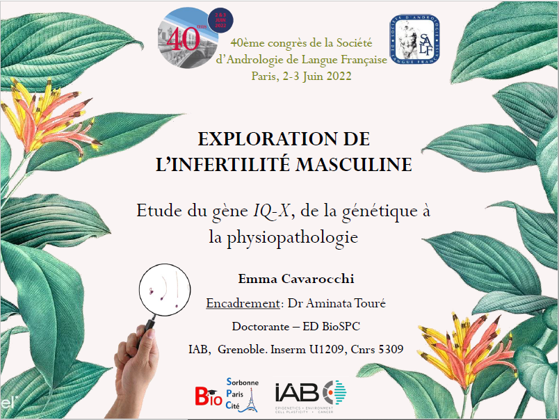
June 2022 - Flash Talk Prize
Congratulations to Emma Cavarocchi who received the Flash Talk prize at the 40th Congress of the French Society of Andrology (SALF) by presenting her latest results on the exploration of the asthenozoospermia phenotype on human and mouse model.

October 2021 - Scientific Prize
Congratulations to Marjorie Whitfield who received the Young Talents prize from the L'Oréal-Unesco Foundation for Women and Science for her work on male reproductive biology and infertility.
No results found.
funding and collaborations
Funding
ANR 'DIVERCIL'
(2017-2022)
Coordinator : B. Durand
ANR 'FLAGELOME'
(2019-2024)
Coordinator : P. Ray
ANR 'SPERMetabo'
(2022-2025)
Coordinator : A. Touré
IRGA 'SLC26 Therapeutics'
(2021-2022)
Coordinator : A. Touré
Individual Fellowship 'SPERMetabo'
(2024-2026)
Coordinator : M. Drouault & A. Touré
France 2030 PUI FITInnovE 'SPERM_NRJ'
(2024-2026)
Coordinator : A. Touré

Bill & Melinda Gates Foundation
(2024)
Coordinator : A. Touré
Core Facilities
Grenoble Alpes University : PHTA Mouse facility, Histology.
Institut Cochin, Paris : Cell Imaging (STORM, STED), Transmission Electron Microscopy.
Institut Cochin, Paris : Protéomique 3P5
Institut Pasteur, Paris : Proteomic.
Bordeaux: University OncoProt.
Toulouse University: Métatoul Lipidomic
National and international collaborations

Pr Bénédicte Durand
Université de Lyon
(France)

Dr Melanie Bonhivers
& Dr Denis Dacheux
Université de Bordeaux
(France)

Dr Anu Sironen
UCL
(UK & Finland)

Dr Zhibing Zhang
Wayne State University
(USA)
Dr Marie-Odile Fauvarque
CEA Grenoble
(France)
Dr Carina Prip-Buus
Institut Cochin
(Paris, France)
Pr Ursula Seidler
Hannover Medical School
(Germany)
Dr Emmanuel Dulioust
& Pr Catherine Patrat
Cochin Hospital
(Paris, France)

Pr Serge Amselem
& Dr Marie Legendre
Trousseau Hospital
(Paris, France)
Dr Feng Zhang
Fudan University
(China)